Epidemiology
Etiology
- Microbiology:
- Staphylococcus aureus: Most common overall (>50%; both MSSA and MRSA).
- Staphylococcus epidermidis: Post-spinal instrumentation, indwelling hardware.
- Escherichia coli & Gram-negative bacilli: Post-UTI, instrumentation of GI/GU tract.
- Pseudomonas aeruginosa & Candida: Injection drug use (IVDU).
- Mycobacterium tuberculosis: Pott disease (endemic areas, immigrants, immunocompromised).
- Brucella: Unpasteurized dairy exposure.
- Routes of Spread:
- Hematogenous seeding (most common): Via arterial supply or retrograde flow through Batson venous plexus.
- Direct inoculation: Spinal surgery, epidural catheterization/injection, LP.
- Contiguous extension: Retropharyngeal abscess, decubitus ulcers, psoas abscess.
- Risk Factors:
- IVDU, DM, ESRD on HD, advanced age, immunosuppression (HIV, chronic steroids).
- Indwelling vascular catheters, recent bacteremia, or spinal procedures.
Pathophysiology
Clinical features
- Vertebral Osteomyelitis / Spondylodiscitis:
- Focal, progressive back pain: Constant, worse at night, and not relieved by rest.
- Point tenderness on percussion over the involved spinous process (high sensitivity).
- Fever present in only ~50% (often absent or low-grade in subacute/chronic cases).
- Limited spinal ROM with surrounding paravertebral muscle spasm.
- Spinal Epidural Abscess (SEA):
- Classic Clinical Triad (present fully in <15%, but high specificity):
- Fever
- Focal spinal back pain (severe, localized)
- Neurologic deficits (radiculopathy → motor/sensory deficits → bowel/bladder incontinence → paralysis)
- Pott Disease (Tuberculous Spondylitis):
- Indolent/chronic course, typically affects lower thoracic/upper lumbar spine.
- Leads to anterior vertebral body destruction, collapse, and gibbus deformity (focal kyphosis).
Diagnostics
- Initial / Screening:
- ESR and CRP: Markedly elevated in >90% of cases; highly sensitive screening tools; used to monitor treatment response.
- CBC: Leukocytosis with left shift (frequently normal in chronic cases).
- Blood Cultures (x2 sets): Positive in up to 50-70%; can identify pathogen and avoid invasive biopsy.
- Confirmatory / Imaging of Choice:
- MRI spine with and without IV contrast: Gold standard (high sensitivity/specificity).
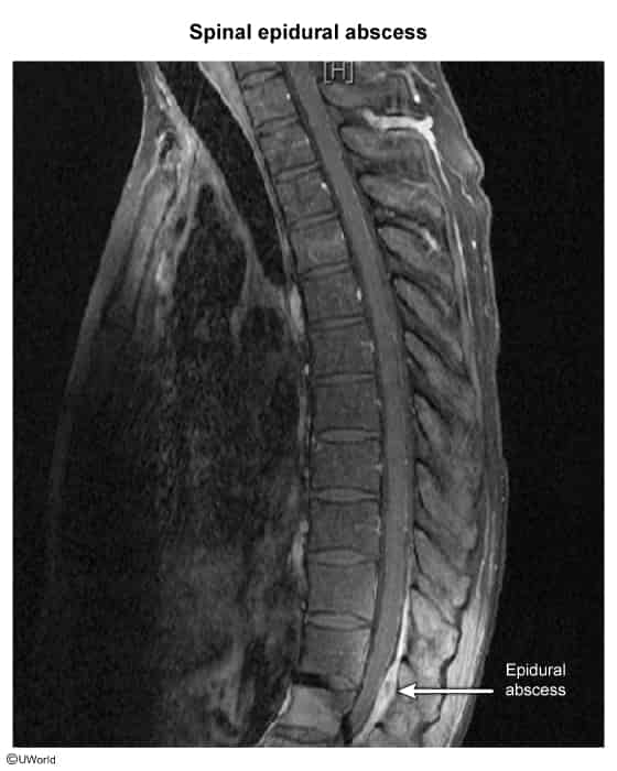
- Shows disc space narrowing, fluid collection, bone marrow edema, and epidural space enhancement.
- Alternative (if MRI contraindicated due to metal/pacemaker): CT spine with IV contrast or CT myelography.
- Plain Radiographs: Low sensitivity in early infection; requires 2-4 weeks to show disc space narrowing and endplate erosion.
- Biopsy / Microbiological Confirmation:
- CT-guided percutaneous needle aspiration/biopsy: Indicated if blood cultures are negative and MRI is abnormal.
- Rule: Hold empiric Abx prior to biopsy unless the patient is hemodynamically unstable or has progressive neurologic deficits.
Treatment