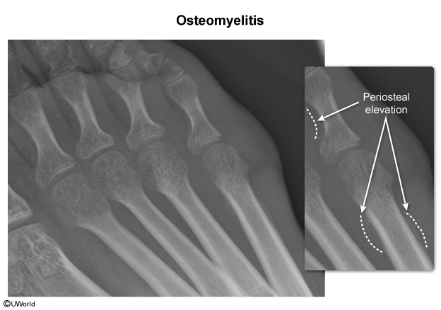
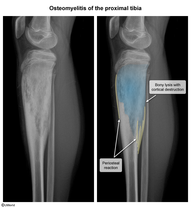
Indolent course: Intermittent, low-grade, deep bone pain with minimal or absent systemic signs (afebrile, normal WBC).
Recurrent sinus tracts: Persistent or intermittent draining cutaneous fistula discharging seropurulent fluid or bone fragments.
Local tissue breakdown: Non-healing ulcer overlying prior trauma/hardware, impaired wound healing, and surrounding indurated/fibrotic skin.
Exposed necrotic bone: Visible, avascular cortical bone or unstable orthopedic implants.
Diagnostics
Initial / Screening Labs: ESR & CRP (highly sensitive; CRP tracks response to Tx). CBC (leukocytosis w/ left shift).
Initial Imaging: Plain Radiographs (X-ray).
May be normal in early disease (first 10-14 days).
Early changes: Soft tissue swelling, periosteal reaction/elevation.
Late changes: Cortical erosion, lytic bone lesions, sequestrum (necrotic bone fragment), involucrum (thick layer of new periosteal bone).
Most Sensitive Imaging: MRI w/ gadolinium (best test for early detection, soft tissue/epidural involvement; indicated if X-ray is negative but clinical suspicion remains high).
Confirmatory / Gold Standard: Bone biopsy and culture (CT-guided needle biopsy or open surgical biopsy).
Must perform before initiating Abx unless pt is hemodynamically unstable or septic.
Superficial wound/sinus tract swabs are unreliable (reflect skin flora; exception: S. aureus).
Treatment
Obtain Diagnostic Samples: Bone biopsy/culture prior to Abx initiation (unless septic/unstable).
Empiric Antibiotics (broad-spectrum, prolonged IV 4-6 weeks):
Standard empiric: Vancomycin (MRSA coverage) + 3rd/4th-gen Cephalosporin (e.g., Cefepime) OR Piperacillin-Tazobactam (Gram-negative & anaerobic coverage).
Sickle cell: Coverage for Salmonella & S. aureus (e.g., Ceftriaxone + Vancomycin). c
Puncture wound (shoe): Coverage for Pseudomonas (e.g., Cefepime or Ciprofloxacin).
Targeted Antibiotics: De-escalate based on bone culture & sensitivity results.
Surgical Intervention: Surgical debridement indicated for sequestrum removal, sinus tract excision, intraosseous/subperiosteal abscess, or failure to respond to Abx.
Monitoring: Serial CRP/ESR levels to track response to therapy.