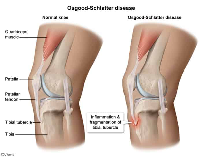
Pathophysiology: Traction apophysitis caused by repetitive strain of the patellar tendon on the immature secondary ossification center of the tibial tuberosity.
Clinical Features
History:
Insidious onset of anterior knee pain.
Exacerbated by running, jumping, squatting, kneeling, and direct trauma.
Relieved by rest.
Physical Examination (PE):
Focal tenderness and prominence/swelling over the tibial tuberosity.
Pain reproduced with resisted knee extension and passive extreme knee flexion.
Normal range of motion (ROM) of the knee joint.
Absence of true knee joint effusion (effusion suggests intra-articular pathology).
Diagnosis
Initial / Primary Approach: Clinical diagnosis based on characteristic history and PE in a growing adolescent.
Imaging (Lateral Knee XR):
Indication: Not routinely required; indicated only for atypical features (e.g., pain at rest, night pain, systemic symptoms, unilateral severe acute pain, soft-tissue mass).
Findings: Soft-tissue swelling over the tibial tuberosity, fragmentation/heterotopic ossification of the secondary ossification center, or irregular apophyseal enlargement.
Confirmatory / Gold Standard: Clinical criteria (routine MRI/CT is not indicated).
Key Labs: Normal (ESR/CRP normal; used only to rule out osteomyelitis, septic arthritis, or malignancy if presentation is atypical).
Differential Diagnostics
Patellar Tendinitis (“Jumper’s Knee”):
Tenderness localized to the inferior pole of the patella (not the tibial tuberosity).
More common in mature/skeletally mature athletes.
Sinding-Larsen-Johansson Syndrome:
Traction apophysitis at the inferior pole of the patella (similar etiology, different anatomical site).
Occurs in slightly younger age group (10–12 y/o).
Tibial Tubercle Avulsion Fracture:
Acute, sudden onset following a violent quadriceps contraction.
Inability to actively extend the knee or bear weight.
Analgesia: Short-term NSAIDs for acute symptom flare-ups.
Physical Therapy: Quadriceps and hamstring stretching and low-impact strengthening.
Protective Equipment: Infrapatellar strap (patellar tendon strap) or protective kneepads.
Second-line (Refractory Symptoms):
Temporary cast or knee immobilizer for 2–4 weeks (reserved strictly for severe, disabling pain failing conservative therapy).
Refractory / Skeletally Mature:
Surgical Excision: Removal of ununited, painful ossicles; only considered after physeal closure (skeletal maturity) if chronic disabling pain persists.
Complications
Permanent cosmetic prominence of the tibial tuberosity (most common).
Chronic localized anterior knee pain due to ununited intratendinous ossicles (<10%).