Patellofemoral pain syndrome
- Epidemiology & Risk Factors:
- Most common cause of anterior knee pain in young female athletes / runners.
- Risk factors: Quadriceps weakness (specifically vastus medialis obliquus [VMO]), hip abductor weakness, increased Q-angle, foot overpronation (pes planus).
- Clinical Presentation:
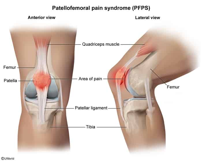
- Poorly localized, dull retropatellar / peripatellar pain provoked by loading a flexed knee (squatting, lunging, stairs).
- “Theater sign” / “Moviegoer’s sign”: Pain/stiffness after prolonged sitting with knees bent.
- No joint effusion; sensation of instability without true mechanical locking.
- Physical Exam:
- (+) Patellar compression test (Clarke test): Pain elicited by compressing the patella into the trochlear groove during active quad contraction.
- Tenderness over patellar facets; lateral patellar tracking during knee extension.
- Diagnosis:
- Clinical diagnosis (initial and best step).
- XR (AP, lateral, sunrise views): Usually normal; rules out OA, fractures, or loose bodies.
- MRI: Not routinely indicated; reserved for refractory cases (>3–6 months) or suspected internal derangement.
- High-Yield Differentials:
- Patellar tendinitis: Focal point tenderness strictly at the inferior patellar pole (jumpers).
- Prepatellar bursitis: Obvious anterior swelling/bursa fluid over the patella; history of repetitive direct kneeling.
- IT band syndrome: Pain localized over the lateral femoral epicondyle; (+) Ober test.
- Osgood-Schlatter: Point tenderness and prominence at the tibial tubercle in growing adolescents.
- Meniscal tear: Joint line tenderness, mechanical locking/catching, joint effusion, (+) McMurray test.
- Management:
- 1st-line: Physical therapy (strengthening quadriceps/VMO and hip abductors/gluteus medius) + activity modification + short-term NSAIDs.
- Adjuncts: Patellar taping/strapping, arch supports/orthotics for pes planus.
- Refractory: MRI evaluation; surgical intervention (e.g., lateral retinacular release) is extremely rare and last-resort.