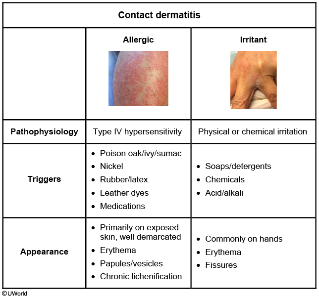
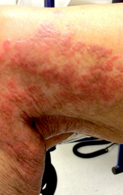
Distribution: Sharply demarcated to the exact areas of direct contact (e.g., hands, web spaces, diaper area sparing inguinal creases).
Acute: Erythema, edema, dryness, cracking, and occasionally vesicles/bullae with caustic agents.
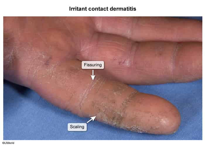
Chronic: Dryness, scaling, hyperkeratosis, painful fissures, and lichenification.
Diagnosis
Initial/Screening: Clinical diagnosis based on detailed occupational/exposure Hx and physical exam findings.
Confirmatory / Rule-Out:
Patch testing: Negative in ICD; used primarily to rule out Type IV hypersensitivity (allergic contact dermatitis).
Key Labs: None routinely indicated.
Biopsy: Rarely needed; reserved for atypical or treatment-refractory cases. Shows non-specific epidermal necrosis, parakeratosis, and superficial perivascular infiltrate.
KOH Prep: Negative for hyphae; performed to exclude dermatophyte infection.
Differential Diagnostics
Allergic Contact Dermatitis (ACD):
Diff: Type IV hypersensitivity (requires prior sensitization); intense pruritus >> burning/pain; lesions typically spread beyond the margins of direct contact; positive patch test.
Dyshidrotic Eczema (Pompholyx):
Diff: Recurrent episodes of intensely pruritic, deep-seated “tapioca-pudding” vesicles on the lateral aspects of fingers, palms, and soles; unrelated to chemical exposure.
Atopic Dermatitis:
Diff: Classic flexural distribution (antecubital and popliteal fossae); personal/family Hx of atopy (asthma, allergic rhinitis); onset typically in early childhood.
Tinea Manuum:
Diff: Often asymmetric/unilateral (classic “two feet, one hand” syndrome); active scaling border with central clearing; positive KOH prep.
Plaque Psoriasis:
Diff: Well-demarcated erythematous plaques with thick silvery scale on extensor surfaces (elbows, knees), scalp, or lumbosacral region; associated with nail pitting and Auspitz sign.
Management
First-Line:
Identify and eliminate exposure to the offending irritant (essential for resolution).
Phototherapy (PUVA or narrowband UVB) for chronic, severe, treatment-resistant occupational disease.
Short course of oral corticosteroids (rarely indicated; reserved for extreme caustic burns).
Complications
Secondary bacterial infection: Most commonly impetiginization (Staph aureus or Strep pyogenes); manifests with honey-colored crusting, purulent discharge, or worsening erythema.
Cellulitis: Entry of pathogens through deep, painful cutaneous fissures.
Chronic disabling dermatitis: Can lead to significant occupational impairment and loss of productivity.
Post-inflammatory pigmentary changes: Hyper- or hypopigmentation, particularly in darker skin phototypes.