Epidemiology & Risk Factors
- Epidemiology: Leading cause of blindness in working-age adults (20–74 yrs).
- Risk Factors: DM duration (strongest predictor), poor glycemic control (↑ HbA1c), HTN, nephropathy, pregnancy.
Clinical Features
- Non-Proliferative Diabetic Retinopathy (NPDR):
- Microaneurysms (earliest sign), dot-and-blot hemorrhages, hard exudates, cotton-wool spots.
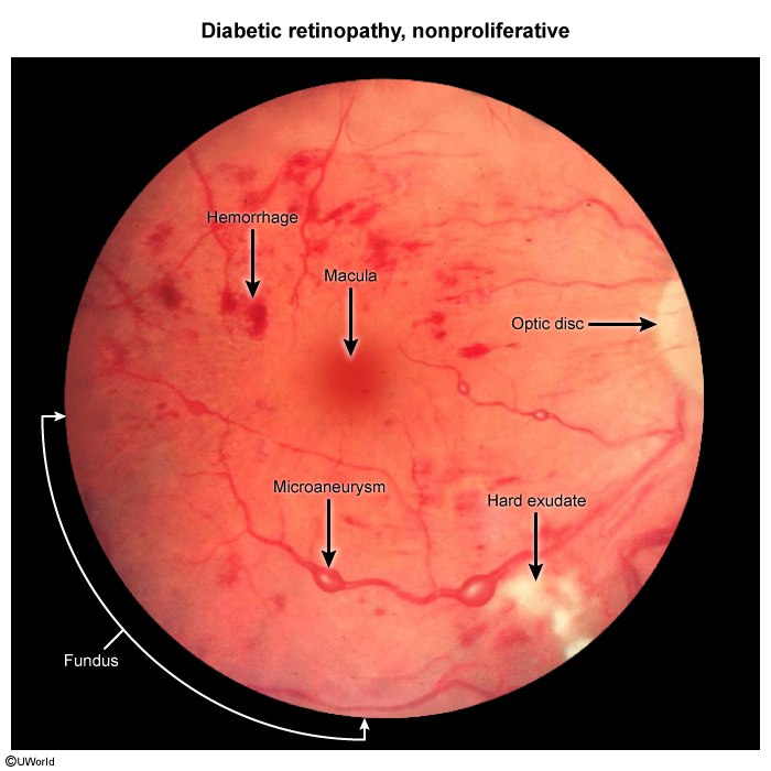
- Macular edema: Leading cause of visual impairment in NPDR.
- Proliferative Diabetic Retinopathy (PDR):
- Driven by retinal ischemia (↑ VEGF).
- Neovascularization (disc/retina/iris), vitreous hemorrhage (sudden floaters/vision loss), tractional retinal detachment.
Diagnosis
- Screening (Dilated Fundoscopy):
- T1DM: 3–5 yrs after dx, then annually.
- T2DM: At time of dx, then annually.
- Confirmatory / Advanced Imaging:
- Optical Coherence Tomography (OCT): Gold standard for macular edema.
- Fluorescein Angiography (FA): Detects non-perfusion and neovascular leakage.
Differential Diagnostics
Management
- Systemic: Strict glucose control (HbA1c ≤ 7.0%) + BP control (< 130/80 mmHg).
- Diabetic Macular Edema: Intravitreal anti-VEGF (e.g., Aflibercept, Bevacizumab).
- Proliferative DR (PDR): Panretinal photocoagulation (PRP) laser and/or anti-VEGF.
- Vitrectomy (Surgery): Non-clearing vitreous hemorrhage, macula-threatening tractional retinal detachment.
Complications
- Vitreous hemorrhage.
- Tractional retinal detachment.
- Neovascular glaucoma.
- Irreversible blindness.