Epidemiology
Etiology
Pathophysiology
- Neoplastic proliferation of plasma cells
- Bone marrow infiltration by malignant plasma cells → suppression of hematopoiesis → leukopenia, thrombocytopenia, anemia
- Cell proliferation → pro-osteoclastogenic factors (e.g., TNF-α, IL-1, RANK-L) → osteolytic lesions → hypercalcemia
- Overproduction of monoclonal immunoglobulin and/or light chains → dysproteinemia (a state of pathologically increased synthesis of immunoglobulins and/or their subunits) → kidney damage (e.g., myeloma cast nephropathy) and/or paraprotein tissue deposition (may cause amyloidosis)
- Nonfunctioning antibodies → functional antibody deficiency
- ↑ Serum viscosity → hyperviscosity syndrome
Warning
Hypercalcemia in MM is not related to PTHrP!
Clinical features
- Often asymptomatic
- Bone pain, especially back pain (most common symptom)
- Symptoms of hypercalcemia, e.g. constipation
- Foamy urine (caused by Bence Jones proteins in urine)
Mnemonic
hyperCalcemia, Renal insufficiency, Anemia, Bone lytic lesions
Diagnostics
- Initial / Screening:
- SPEP (Serum Protein Electrophoresis) & UPEP (Urine Protein Electrophoresis) with Immunofixation: Demonstrates monoclonal paraprotein spike (M-spike).
- Serum Free Light Chain (SFLC) Assay: Quantifies kappa/lambda light chain ratio (highly sensitive).
- Urinalysis vs. 24-hr Urine Total Protein:
- Standard U/A dipstick is negative or trace for protein (dipstick detects only albumin, not Bence-Jones light chains).
- 24-hr urine protein is markedly elevated (detects Bence-Jones proteins).
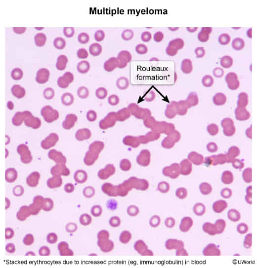
- Peripheral Blood Smear: Rouleaux formation (RBC stacking due to elevated serum paraprotein neutralizing negative RBC charges).
- Key Labs:
- CBC: Normocytic, normochromic anemia.
- Chemistry: ↑ Ca²⁺, ↑ BUN/Cr, normal or low albumin with elevated total protein (“protein-albumin gap” > 4 g/dL).
- Inflammatory Markers: Markedly elevated ESR (> 100 mm/hr).
- Prognostic Labs: Elevated β2-microglobulin (correlates with tumor burden) and LDH.
- Imaging:
- Whole-body low-dose CT (WBLDCT) or Whole-body PET/CT / MRI: Modality of choice for detecting focal bone lesions.
- Skeletal Survey (Plain X-rays): Demonstrates classic “punched-out” osteolytic lesions without surrounding sclerosis, diffuse osteopenia, or pathologic fractures.
- High-Yield Pearl: Technetium-99m bone scan is contraindicated / unreliable (bone scans detect osteoblastic activity; MM lesions are purely osteolytic, causing false-negative scans).
Bone marrow biopsy
- Cytology: clusters of plasma cells
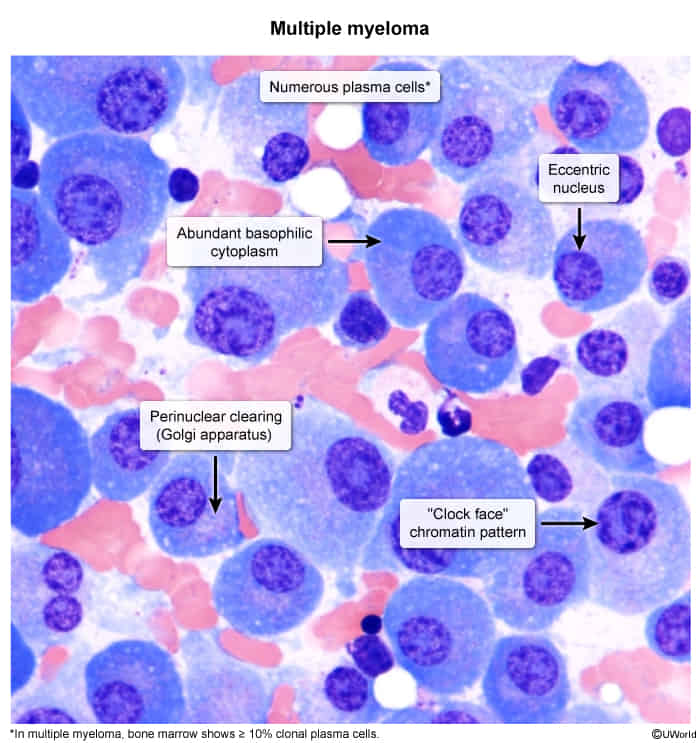
- Mildly organized monoclonal cells
- Perinuclear lucent zone
- Active Golgi apparatus
- Clockface nuclei: Chromatin in the periphery of the nucleus resembles a cartwheel or clock face arrangement.
- Intracytoplasmic crystalline inclusion bodies containing IgG
Differential diagnostics
Waldenström macroglobulinemia
- Pathophysiology: Clonal B-cell/lymphoplasmacytic neoplasm producing excess monoclonal IgM; associated with MYD88 L265P mutation (>90%).
- Clinical Presentation:
- Hyperviscosity Syndrome:
- Neurologic: Headache, dizziness, confusion, ataxia, stroke-like symptoms.
- Visual: Blurry vision; fundoscopy shows dilated, tortuous “sausage-link” retinal veins and retinal hemorrhages. c
- Bleeding: Epistaxis, gingival oozing (IgM impairs platelet function and clotting factors).
- Lymphadenopathy (LAD) & Hepatosplenomegaly (HSM) (present in ~20–30%; distinguishes from Multiple Myeloma).
- Other Findings: Sensorimotor peripheral neuropathy, cold agglutinin disease / cryoglobulinemia (Raynaud, livedo reticularis), B-symptoms (fever, night sweats, weight loss), normocytic anemia.
- Key Negative: No osteolytic bone lesions and no hypercalcemia (unlike Multiple Myeloma).
- Hyperviscosity Syndrome:
- Diagnosis:
- Initial: SPEP + Immunofixation (IFE) revealing monoclonal IgM M-spike.
- Peripheral Blood Smear: Rouleaux formation.
- Confirmatory (Gold Standard): Bone marrow biopsy demonstrating ≥10% clonal lymphoplasmacytic infiltration.
- Key Differentials:
- Multiple Myeloma (MM): CRAB criteria (HyperCalcemia, Renal failure, Anemia, Bone lytic lesions), IgG/IgA spike (rarely IgM), pure plasma cells on BM, no LAD/HSM.
- IgM MGUS: Asymptomatic, IgM <3 g/dL, BM clonal cells <10%, no hyperviscosity or end-organ damage.
- Management:
- Asymptomatic: Observation / Watch-and-wait.
- Acute Hyperviscosity (Emergency): Emergent Plasmapheresis (PLEX) to rapidly decrease intravascular IgM.
- Caution: Do not transfuse packed RBCs before PLEX (increases blood viscosity and worsens symptoms).
- First-line Systemic Therapy:
- Rituximab-based regimens (e.g., Rituximab + Bendamustine). Must perform PLEX before Rituximab if IgM is markedly elevated to prevent a fatal “IgM flare”.
- BTK inhibitors (e.g., Ibrutinib, Zanubrutinib).
Treatment
Complications
- Myeloma cast nephropathy (i.e., myeloma kidney): most common cause of renal injury and renal failure in patients with multiple myeloma
- Clinical features: oliguria, peripheral edema, dyspnea
- Pathophysiology: excessive production and filtration of light chains into the urine → precipitation of light chains in renal tubules → tubular obstruction
- Diagnosis: markedly positive urine sulfosalicylic acid test and/or urine protein electrophoresis
- On light microscopy, numerous large, glassy eosinophilic casts are seen.
- May progress to end-stage renal disease (ESRD)
- AL amyloidosis: Light chains can accumulate as amyloids and may lead to restrictive cardiomyopathy, renal insufficiency, macroglossia, and malabsorption syndromes.
- Infections
- Secondary plasma cell leukemia