Small intestine
Duodenum
- Brunner glands
- Lined by columnar cells that secrete mucus and HCO3−
- Secretions neutralize acidic chyme from the stomach.
- Meissner plexus
Microscopic anatomy
- General Architectural Features
- Mucosal surface area is dramatically increased for absorption via:
- Plicae circulares (Valves of Kerckring): Macroscopic folds of mucosa and submucosa. Most prominent in jejunum.
- Villi: Finger-like projections of the mucosa.
- Microvilli: Form the brush border on the apical surface of enterocytes.
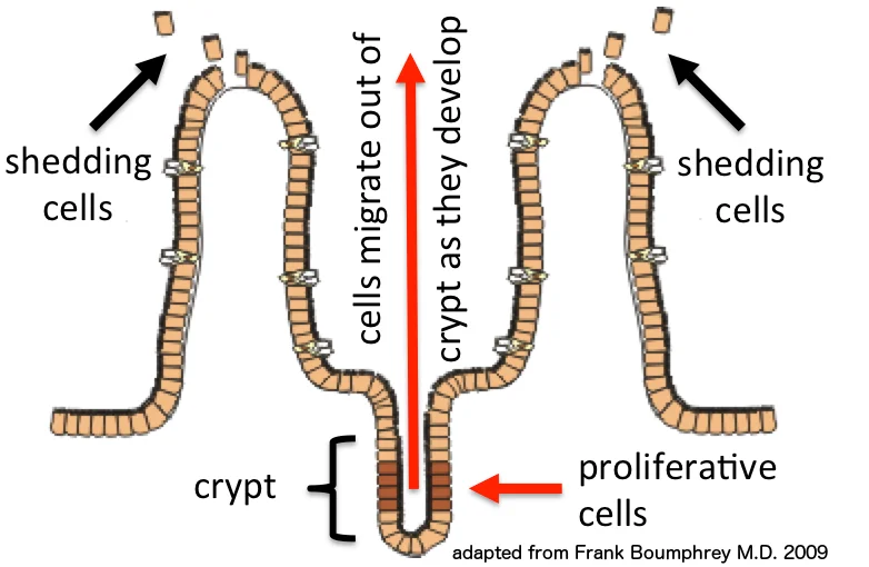
- Crypts of Lieberkühn: Tubular glands located between villi. Contain stem cells and Paneth cells.
- Key Cell Types
- Enterocytes: Primary absorptive cells. Tall columnar with apical microvilli.
- Goblet Cells: Secrete mucin for lubrication and protection. Number increases distally (Duodenum < Jejunum < Ileum).
- Paneth Cells: Located at the base of crypts. Secrete antimicrobial substances (lysozyme, defensins).
- Enteroendocrine Cells: Secrete hormones (e.g., CCK, secretin, GIP).
- M (Microfold) Cells: Specialized cells overlying Peyer’s patches that transport antigens.
- Duodenum
- Distinguishing Feature: Brunner’s Glands in the submucosa (secrete alkaline mucus).
- Villi: Leaf-shaped.
- Jejunum
- Distinguishing Feature: Longest, finger-like villi and most prominent plicae circulares.
- Function: Maximum absorption.
- Ileum
- Distinguishing Feature: Peyer’s Patches in the submucosa.
- Function: Absorbs Vitamin B12 and bile salts.
- Histology: Most numerous goblet cells; shortest, club-shaped villi.