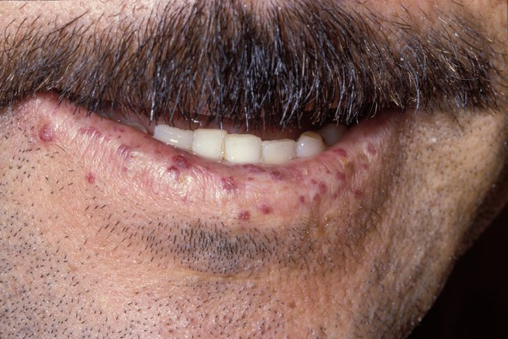
Etiology & Genetics: Autosomal dominant (AD) vascular dysplasia leading to localized loss of capillary beds and widespread direct arteriovenous malformations (AVMs).
Key Clinical Features (Curaçao Criteria):
Recurrent spontaneous epistaxis: Most common initial presentation. c
Mucocutaneous telangiectasias: Small, blanching red macules on lips, tongue, oral mucosa, and fingertips.
Visceral AVMs:
Pulmonary AVMs: R-to-L shunt → cyanosis, digital clubbing, reactive polycythemia, and hemoptysis. c
GI AVMs: Occult bleeding → severe iron deficiency anemia (IDA).
Hepatic AVMs: Can cause high-output heart failure (HF).
Brain abscess: Caused by bacteremia bypassing lung capillary filtration via pulmonary AVMs.
Paradoxical embolic stroke: DVT traversing pulmonary AVM to cerebral circulation.
Diagnostic Workup:
Pulmonary AVM screening (Initial): Contrast echocardiography (“bubble study”) showing delayed appearance of bubbles in the left atrium (>3–8 cardiac cycles).
Pulmonary AVM confirmation: Chest HRCT.
Cerebral AVM screening: Brain MRI with contrast.
Labs: Microcytic anemia (bleeding) OR polycythemia (hypoxemia from R-to-L shunt).
High-Yield Management:
Pulmonary AVMs: Transcatheter embolization (coiling) to prevent stroke and brain abscess.
Infection prevention: Antibiotic prophylaxis prior to dental/surgical procedures.
Symptom & Bleeding control: Nasal humidification/cautery, iron repletion, and Bevacizumab (anti-VEGF) for refractory bleeding.