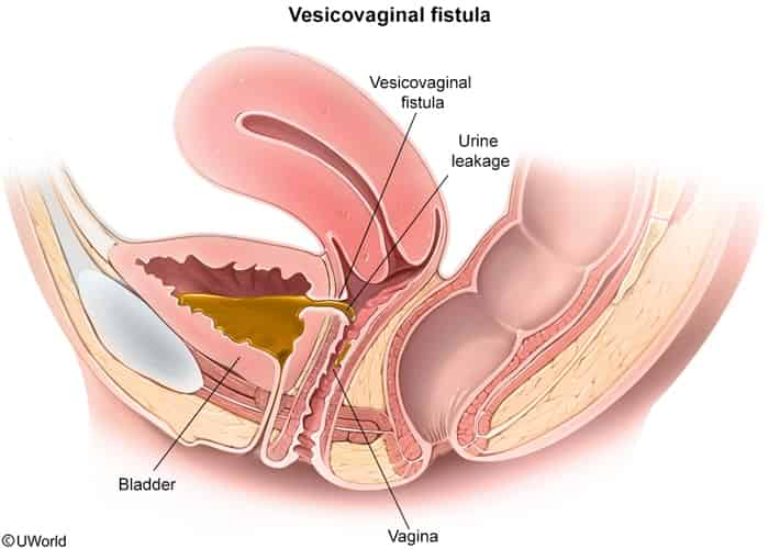
Developing Nations: Obstructed labor (most common cause worldwide; pressure of fetal head causes tissue ischemia and necrosis).
Developed Nations: Gynecologic surgery (most common cause; especially Total Abdominal Hysterectomy [TAH]), pelvic radiation therapy, pelvic malignancy, IBD (Crohn disease), or obstetric trauma.
Anatomic Types:
Vesicovaginal Fistula (VVF): Most common type overall.
Ureterovaginal Fistula: Associated with ureteral injury during lower abdominal/pelvic surgery.
Urethrovaginal Fistula: Linked to urethral diverticulum surgery or prolonged labor.
Clinical Features
Continuous, painless leakage of urine from the vagina (classic presentation 1–2 weeks post-pelvic surgery or delivery).
Preserved normal voiding pattern: Pts may still void normally if the fistula is small or if it is a unilateral ureterovaginal fistula.
Perineal skin breakdown: Erythema, excoriation, and dermatitis secondary to chronic moisture.
Vaginal discharge: Malodorous, watery discharge; recurrent UTIs or pyelonephritis.
Physical Exam: Speculum exam shows pooling of fluid in the posterior vaginal fornix or a visible defect.
Diagnosis
Initial Test / Bedside Evaluation:
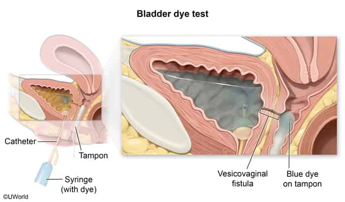
Retrograde Dye Test (Methylene Blue / Indigo Carmine Test):
Instill blue dye into the bladder via Foley catheter while placing a vaginal tampon.
Tampon turns blue: Diagnosis of Vesicovaginal Fistula (VVF) confirmed.
Tampon stays clear, but wet w/ urine: Suggests Ureterovaginal Fistula (dye bypassed because leakage originates from upper urinary tract).
Conservative Management (First-line for small VVF <0.5 cm detected early post-op):
Continuous Bladder Drainage via Foley Catheter for 4–6 weeks.
Allows small, fresh tracts to heal spontaneously without surgical intervention.
Surgical Management (Definitive / Refractory or Large VVF):
Timing: Delayed repair 2–3 months (up to 6 months post-radiation/inflammation) after initial injury to allow surrounding tissue inflammation and edema to resolve.
Surgical Approaches: Transvaginal or transabdominal excision of fistula tract w/ layered closure and interposition flaps (e.g., Martius fat pad flap).
Ureterovaginal Fistula Management: Ureteral stenting (if minor/early) or Ureteroneocystostomy (ureteral reimplantation into the bladder).
Complications
Recurrent UTIs and chronic pyelonephritis.
Ureteral stricture, hydronephrosis, and permanent renal failure.
Vaginal stenosis and dyspareunia.
Chronic perineal dermatitis and vulvar excoriation.
High rate of fistula recurrence if repaired under inflamed or radiated conditions.