Etiology


  • Maternal risk factors
    • Folate deficiency during pregnancy due to:
      • Insufficient folate supplementation
      • Drugs that interfere with folate metabolism
  • Pregestational diabetes mellitus

Closed spinal dysraphism


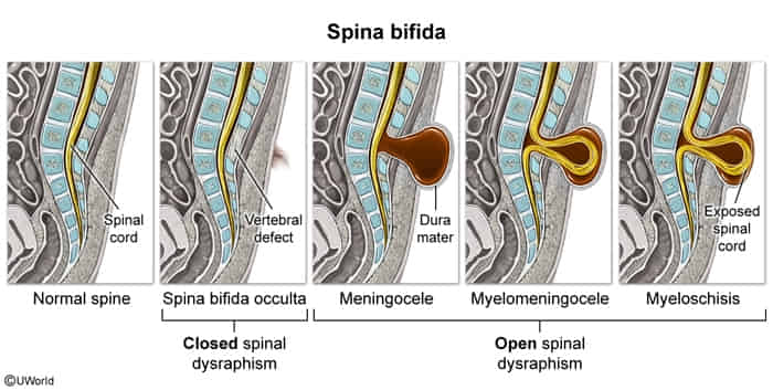
Spina bifida occulta

  • Description
    • Most common closed NTD
    • Cause: failure of vertebral arch fusion
    • Vertebral bone defect without herniation
    • The spinal cord, meninges, and overlying skin remain intact.
  • Clinical features
    • Most commonly affects the lower lumbar or sacral region
    • Often asymptomatic (may be an incidental finding in imaging)
    • Possible symptoms at the level of the vertebral defect:
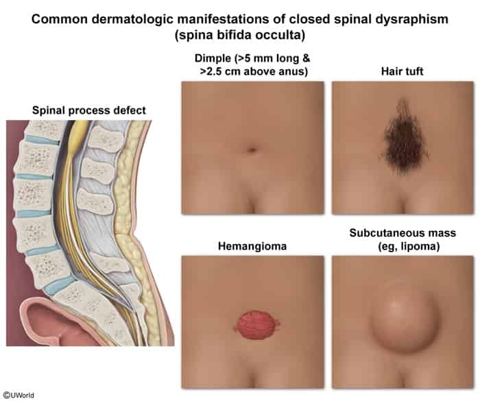
      • Lumbar skin dimple
      • Collection of fat
      • Patch of hair
      • Vascular malformations (hemangioma, port-wine stain).
  • Diagnostics

Diagnostics


Prenatal period

  • Screening test (16–18 weeks’ gestation): AFP in maternal serum (MSAFP)
    • MSAFP is only elevated in open NTDs.
      • Because the fetal tissues (which are producing AFP) are directly exposed to the amniotic fluid, there is a significantly increased leakage of AFP from the fetus into the amniotic fluid.
    • MSAFP is not elevated in spina bifida occulta.
  • Ultrasonography (18–20 weeks’ gestation)
    • Characteristic findings depend on the specific defect.
      • Findings in anencephaly
        • Cranial vault and brain tissue are absent.
        • Residual, disorganized cerebellar and/or brainstem tissue may be present.
        • Bulging eyes and underdeveloped forehead
        • Associated with polyhydramnios
  • Amniocentesis: ↑ AFP and ↑ AChE in amniotic fluid (increase in open NTDs only)
    • During fetal development, AChE is present in the developing nervous system tissues, including the neural tube.
    • Used as confirmation test when MSAFP is elevated but ultrasound findings are inconclusive
    • When both AFP and AChE are elevated, an open NTD is very likely.

Differential diagnostics

Tethered cord syndrome

  • Definition & Pathophysiology:
    • Spinal cord stretch/traction injury due to mechanical anchoring of the conus medullaris below L1–L2 (thickened filum terminale, lipoma, spina bifida occulta).
    • Strongly associated with spina bifida occulta and other forms of closed spinal dysraphism.
    • Exacerbated during growth spurts (age 5–15) or spinal flexion.
  • Clinical Presentation:
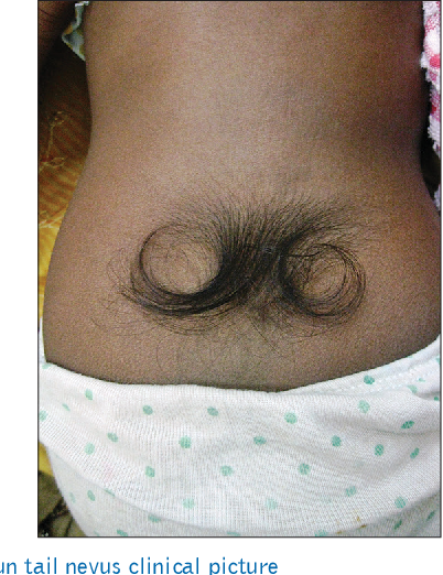
    • Cutaneous stigmata (lumbosacral midline): “Faun tail” (tuft of hair), sacral dimple (>0.5 cm wide, >2.5 cm above anal verge), subcutaneous lipoma, or hemangioma.
    • Neuromusculoskeletal: Progressive LE weakness, gait instability, mixed UMN/LMN signs, progressive scoliosis, and foot deformities (pes cavus, hammer toes). c
    • Urologic / GI: Neurogenic bladder (new-onset incontinence, daytime wetting, recurrent UTIs), chronic constipation, encopresis.
  • Diagnosis:
    • Initial (< 4–6 months old): Spinal Ultrasound (viable prior to posterior arch ossification).
    • Confirmatory (> 6 months old & Gold Standard): Lumbosacral MRI (demonstrates low-lying conus < L2 and thickened filum > 2 mm).
    • Ancillary: Urodynamic studies (assesses detrusor hyperreflexia/sphincter dyssynergia).
  • Key Differentials:
    • Cauda equina syndrome: Acute onset, massive disc herniation, saddle anesthesia, absent congenital skin stigmata.
    • Charcot-Marie-Tooth (CMT): Distal hereditary polyneuropathy (“inverted champagne bottle” legs), normal spine imaging.
  • Management:
    • Definitive: Early surgical detethering (laminectomy w/ filum terminale transection) to stop progressive irreversible neuro/urologic loss.
    • Supportive: Clean intermittent catheterization (CIC), anticholinergics (oxybutynin), PT/OT, corrective orthotics/surgery.
  • Complications:
    • Irreversible neurogenic bladder → hydronephrosis → chronic kidney disease (CKD).
    • Permanent paraparesis and postoperative re-tethering from scar tissue.