- Definition: benign capillary vascular tumor of infancy
- Epidemiology
- Occurs in 3–10% of infants
- More commonly affects girls
- Pathophysiology
- Abnormal development of vascular endothelial cells
- Rapid proliferation followed by subsequent spontaneous slow involution (occurring at the age of 5–8 years)
- Spontaneous resolution is common
- Clinical course:
- Absent/faint at birth → rapid proliferation 3–9 mo → spontaneous involution (50% by age 5, 90% by age 9).
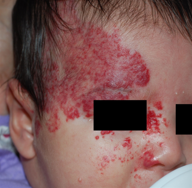
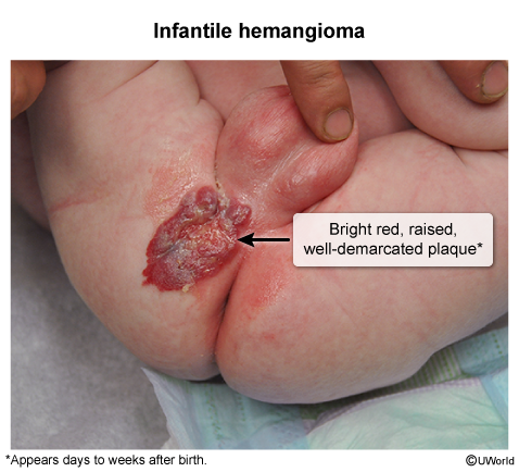
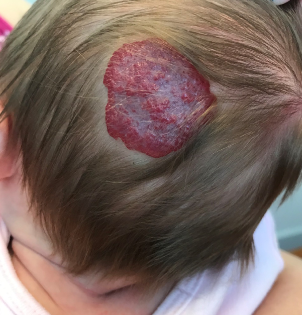
- Superficial = bright red “strawberry” plaque; deep = bluish, doughy subcutaneous mass.
- High-risk locations:
- Periocular → visual axis obstruction, amblyopia, astigmatism.
- “Beard” area → subglottic/airway hemangioma (biphasic stridor).
- Lumbosacral → spinal dysraphism/tethered cord.
- ≥5 cutaneous lesions → hepatic hemangiomas (high-output HF).
- Segmental facial >5 cm → PHACE syndrome.
- DDx:
- Port-wine stain: present at birth, grows proportionally, never involutes, GLUT-1 (−).
- Congenital hemangioma (RICH/NICH): fully formed at birth, GLUT-1 (−).
- Pyogenic granuloma: older child, friable, bleeds with trauma.
- Kaposiform hemangioendothelioma: purpuric plaque + Kasabach-Merritt (consumptive thrombocytopenia).
- Nevus simplex: flat, midline, fades spontaneously.
- Management:
- Uncomplicated → observation + reassurance (most involute).
- High-risk (vision/airway compromise, ulceration, disfigurement, HF) → oral propranolol (1st line; monitor hypoglycemia, bradycardia, hypotension). c
- Small superficial lesion → topical timolol.
- Propranolol CI/failure → systemic corticosteroids.
- Refractory → pulsed-dye laser (ulceration/telangiectasias), surgical excision.