Epidemiology
Etiology
- Prior breast trauma (e.g., seatbelt injury, sports injury; up to 50% may not recall specific trauma).
- Iatrogenic breast injury: Recent surgery (e.g., lumpectomy, reduction, augmentation, reconstruction), core needle biopsy, or radiation therapy.
- More common in obese women with large, pendulous breasts.
- Strictly benign non-proliferative breast lesion; no increased risk of breast carcinoma.
Pathophysiology
Dead fat tissues replaced by scar tissues
Clinical features
- Palpable breast mass: Typically firm, irregular, fixed or mobile, painless or mildly tender.
- Skin changes: May present with skin dimpling, tethering, nipple retraction, or overlying ecchymosis/erythema (frequently mimics breast carcinoma on PE).
- Systemic signs (fever, leukocytosis) and axillary lymphadenopathy are usually absent.
Diagnostics
- Initial Evaluation: Diagnostic imaging tailored by age (age < 30: Targeted US; age ≥ 30: Diagnostic Mammography + Targeted US).
- Mammography: Lucent area (fat density), “oil cyst” with eggshell/rim calcifications, coarse dystrophic calcifications, or an irregular spiculated mass. c
- Ultrasound: Anechoic or hypoechoic mass, complex cystic lesion with internal echoes/mural nodules, or shadowing mass.
- Confirmatory / Gold Standard: Core Needle Biopsy (CNB) (indicated whenever imaging findings are indeterminate or suspicious for malignancy [BI-RADS 4/5]).
- Histopathology: c
- Foamy lipid-laden macrophages (histiocytes).
- Multinucleated giant cells, fat globules/vacuoles, and hemosiderin deposition.
- Chronic inflammatory infiltrate and peripheral fibrosis.
Mnemonic
Cheese
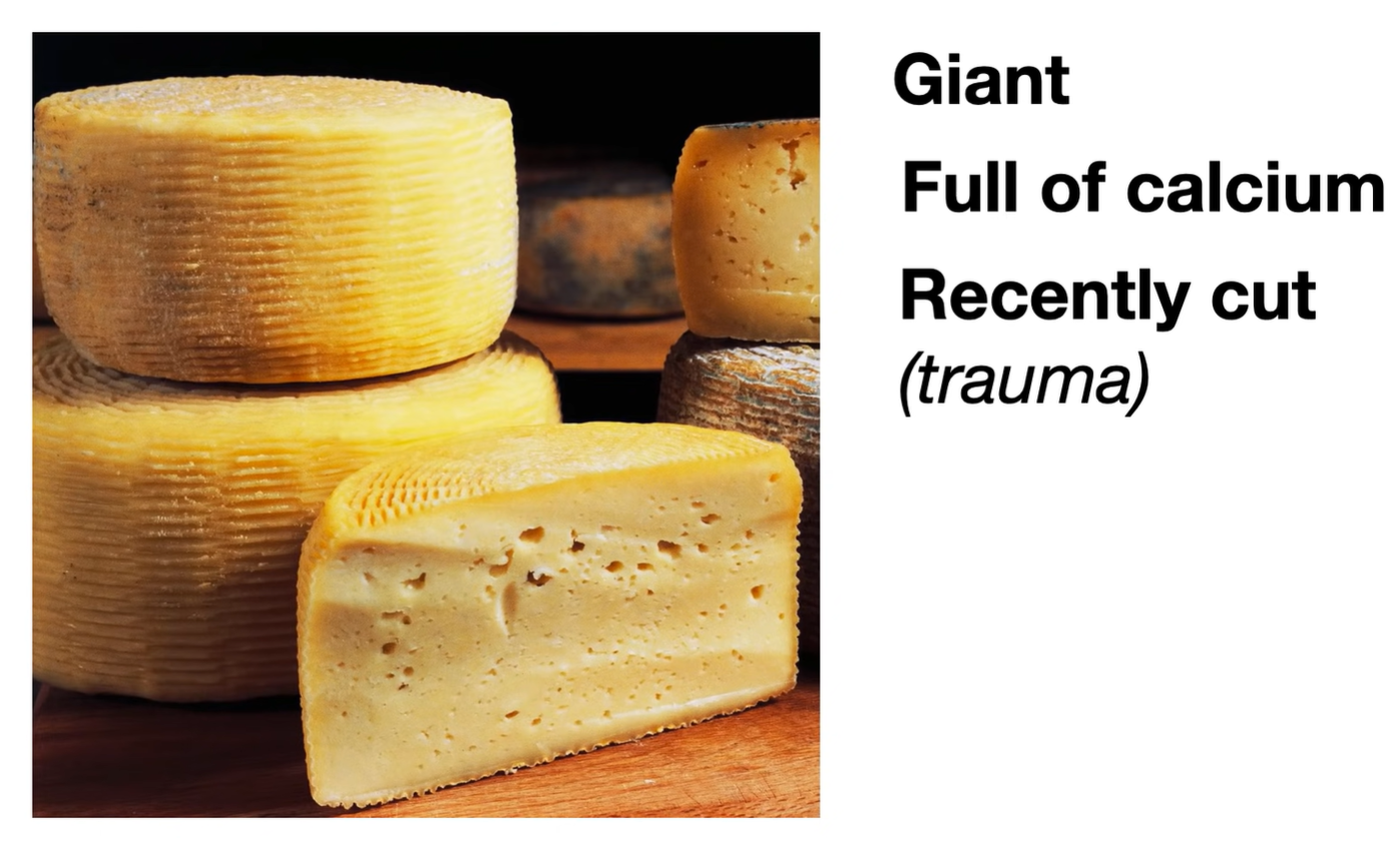
Differential Diagnostics
- Invasive Ductal Carcinoma (IDC):
- Differentiating feature: Both present as fixed, hard, irregular masses with skin retraction. IDC lacks benign histopathology and shows malignant epithelial cells invading stroma; mammogram shows pleomorphic microcalcifications/spiculation.
- Fibroadenoma:
- Differentiating feature: Well-circumscribed, highly mobile, non-tender, rubbery mass in young females (< 35 yo); no history of trauma/surgery; US shows oval, well-demarcated mass with uniform hypoechogenicity.
- Fibrocystic Changes:
- Differentiating feature: Bilateral, diffuse, cyclical breast pain/lumpiness that fluctuates with the menstrual cycle; multiple small cysts on US.
- Breast Abscess:
- Differentiating feature: Presents with acute inflammatory signs (erythema, warmth, severe pain, fluctuance, fever); primarily seen in lactating females (mastitis progression).
- Galactocele:
- Differentiating feature: Subareolar, non-tender, cystic mass occurring during or shortly after cessation of lactation; aspiration reveals milky fluid.
Treatment
- First-line (Observation & Reassurance):
- Reassurance once malignancy is definitively ruled out via imaging ± CNB.
- Lesions typically resolve spontaneously or persist as stable, asymptomatic oil cysts/calcifications over months to years.
- Symptomatic Management:
- NSAIDs and supportive bra for localized breast tenderness/discomfort.
- Procedural / Surgical (Refractory / Uncertain):
- Needle Aspiration: For symptomatic, tense, painful oil cysts.
- Surgical Excision: Reserved for persistent diagnostic ambiguity, non-resolving painful masses, or growing lesions concerning for occult malignancy.