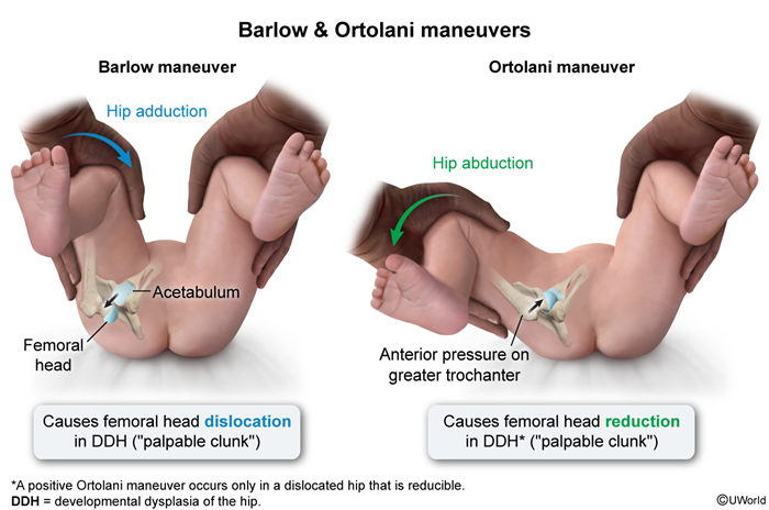
Limited hip abduction (< 60° while supine with hips flexed at 90°).
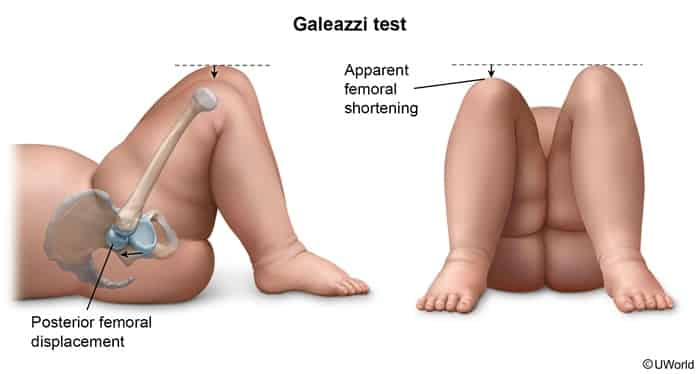
Galeazzi sign: Asymmetry in knee height with hips and knees flexed (femur appears shortened on the affected side).
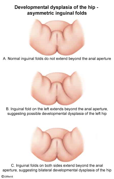
Asymmetric skin folds (inguinal/thigh/gluteal; sensitive but non-specific).
Toddlers / Walking Age:
Trendelenburg gait (unilateral DDH: pelvis tilts toward unaffected side during stance on affected leg).
Waddling gait (bilateral DDH).
Leg length discrepancy (LLD) and compensatory toe-walking.
Diagnosis
Initial / Screening:
Serial physical exams (Barlow & Ortolani) at every well-child visit until walking age.
Imaging Modality Choice (Age-Dependent):
Age < 4–6 months: Hip Ultrasonography (cartilaginous unossified femoral head; dynamic stress testing reveals displacement and measured α-angle <60∘).
Perform US at 4–6 weeks of life (not at birth) to avoid false positives from physiologic ligamentous laxity.
Age ≥ 4–6 months: AP and Frog-leg Pelvic Radiographs (ossification center of femoral head appears; assess acetabular index, Perkins line, and Shenton line disruption).
Confirmatory:
Abnormal US (α-angle <60∘ or coverage <50%) or abnormal pelvic X-ray.
Differential Diagnostics
Transient Synovitis: Diff by acute-onset limp/hip pain following a viral URI in children aged 3–8 yrs; normal bony anatomy on imaging.
Septic Arthritis: Diff by high fever, refusal to bear weight, elevated inflammatory markers (ESR/CRP), joint effusion, and synovial WBC >50,000/μL.
Legg-Calvé-Perthes Disease (LCPD): Diff by idiopathic avascular necrosis in boys aged 4–10 yrs; presents with insidious limp and pain; X-ray shows femoral head collapse/flattening.
Slipped Capital Femoral Epiphysis (SCFE): Diff by obese adolescent (10–16 yrs) with dull groin/thigh/knee pain and limited internal rotation; X-ray shows posterior displacement of femoral head (“ice cream slipping off cone”).
Benign Hip Clicks: Diff by high-pitched, soft fascial snap with normal range of motion, negative Barlow/Ortolani maneuvers, and normal US.
Management
Age < 6 months:
First-line: Pavlik harness (dynamic splint maintaining hip in flexion and abduction for 6–12 weeks).
Monitor closely via weekly/bi-weekly US to confirm reduction and avoid complications.
Age 6–18 months (or failed Pavlik):
Closed reduction under general anesthesia followed by hip Spica cast immobilization (typically for 3 months).
Age > 18 months (or failed closed reduction):
Open surgical reduction± pelvic/femoral osteotomy + postoperative Spica cast.
Complications
Avascular Necrosis (AVN) of the femoral head (most feared complication of Pavlik harness if placed in hyperabduction/hyperflexion or after aggressive reduction).
Femoral nerve palsy (transient; caused by excessive hip flexion in harness).
Early-onset hip osteoarthritis (secondary to residual acetabular dysplasia).
Persistent limb length discrepancy and gait abnormalities.