Neonatal soft tissue injuries
Soft tissue injuries of the scalp in infants are mostly caused by shearing forces during vacuum or forceps delivery.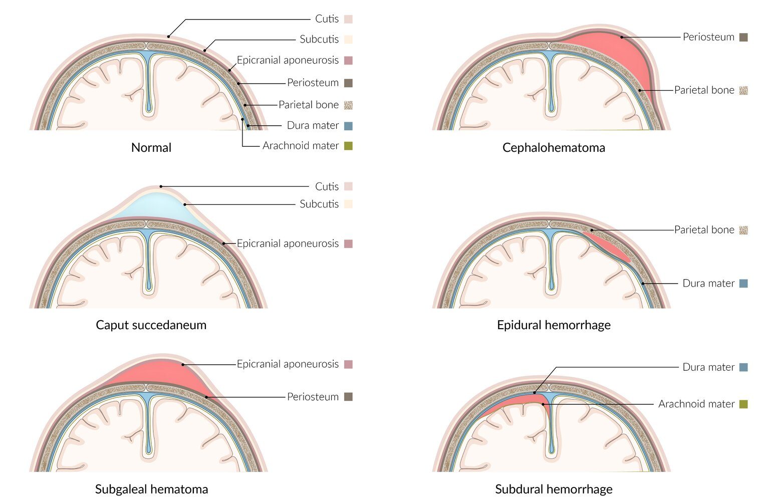
Epidemiology & Risk Factors
- Operative vaginal delivery: Vacuum extraction (highest risk for subgaleal hemorrhage), forceps delivery.
- Fetal factors: Macrosomia / Large for gestational age (LGA), infant of diabetic mother (IDM), abnormal presentation (face, brow, breech), shoulder dystocia.
- Maternal/Labor factors: Primiparity, prolonged second stage of labor, precipitous delivery, cephalopelvic disproportion (CPD).
- Iatrogenic: Fetal scalp electrode (FSE) placement, intrauterine pressure catheter (IUPC).
Clinical Features
- Caput Succedaneum:
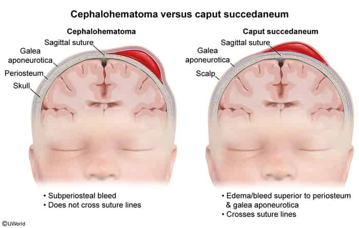
- Subcutaneous serosanguinous fluid accumulation superficial to the periosteum.
- Crosses suture lines, soft, pitting, poorly demarcated margins. c
- Present at delivery; resolves spontaneously within 2–3 days.
- Cephalohematoma:
- Subperiosteal hemorrhage (rupture of subperiosteal blood vessels).
- Does NOT cross suture lines (limited by cranial sutures/periosteum).
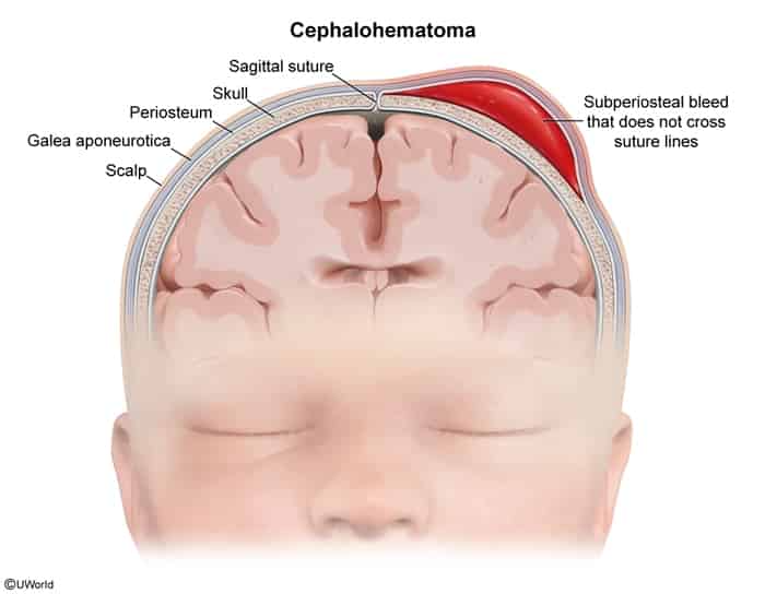
- Firm, fluctuant with well-demarcated edges; may enlarge over hours to days. c
- Resolves over weeks to months; may undergo central liquefaction or peripheral calcification (“eggshell” rim).
- Subgaleal Hemorrhage (Emergent): c
- Rupture of emissary veins leading to blood accumulation in the loose connective tissue between the epicranial aponeurosis (galea) and periosteum.
- Crosses suture lines and cranial ridges; diffuse, fluctuant, progressive swelling spreading across the entire scalp, neck, and behind ears (ecchymosis).
- Systemic signs: Tachycardia, hypotension, pallor, rapid drop in hematocrit, lethargy, expanding head circumference (1 cm increase ≈ 30–40 mL of blood loss).
- Congenital Muscular Torticollis (Fibromatosis Colli):
- SCM injury/hematoma with fibrous contracture due to intrauterine crowding or birth trauma.
- Palpable, nontender, mobile SCM mass/olive noted at 2–4 weeks of life.
- Head tilted toward affected SCM and chin rotated away to the contralateral side.
- Subcutaneous Fat Necrosis:
- Firm, non-tender, violaceous/erythematous nodules or plaques on the back, buttocks, cheeks, or shoulders.
- Occurs days-to-weeks after birth trauma, asphyxia, or therapeutic hypothermia.
Birth-related clavicle fracture
- Epidemiology & Risk Factors: Most common birth-related fracture; associated with fetal macrosomia (birth weight > 4000 g, maternal GDM), shoulder dystocia, and operative vaginal delivery (forceps/vacuum).
- Clinical Presentation:
- Localized crepitus, swelling, or bony step-off over the clavicle.
- Asymmetric Moro reflex with normal grasp reflex.
- Pseudoparalysis (decreased spontaneous arm movement due to pain, not true motor deficit).
- Palpable, nontender callus formation at 1–3 weeks (reassure parents this is normal healing).
- Diagnosis:
- Initial & Confirmatory: Clavicle XR (identifies fracture site and rules out concomitant humeral fracture).
- PE: Rule out concurrent brachial plexus injury (e.g., Erb-Duchenne palsy: C5–C6, “waiter’s tip” posture, absent biceps reflex).
- Management:
- First-line: Reassurance and gentle handling; complete spontaneous healing expected in 2–4 weeks without long-term sequelae. c
- Immobilization: Pin sleeve to shirt with elbow flexed at 90° for comfort (rigid splints, casts, or figure-of-8 bandages are not recommended).
- Analgesia: Acetaminophen prn for the first few days.
- Complications: Rare (transient brachial plexus neuropraxia; pneumothorax or subclavian vessel injury is extremely rare).