Infectious
Postpartum endometritis
Definition & Etiology :
Most common cause of postpartum fever (day 2–3).
Polymicrobial: Vaginal flora (anaerobes: Bacteroides , Peptostreptococcus ; aerobes: E. coli , GBS).
Risk Factors :
Cesarean delivery (single most important risk factor).PROM (>18 hrs) , prolonged labor, operative vaginal delivery, chorioamnionitis.
Clinical Features :
Postpartum fever (≥38.0°C / 100.4°F) on day 2–3 .
Foul-smelling / purulent lochia .Uterine / fundal tenderness , lower abdominal pain, leukocytosis.
Diagnosis :
Clinical : Fever + uterine tenderness + foul lochia post-delivery.Pelvic US : Indicated only if refractory to Abx to rule out RPOC or pelvic abscess .Endometrial cultures not routinely recommended (contamination risk).
Management :
1st-line : IV Clindamycin + IV Gentamicin until afebrile x 24–48 hrs (add Ampicillin if non-responsive or Enterococcus suspected). c Refractory (>48–72 hrs Abx) :
Pelvic US : Check for RPOC (treat w/ D&C ) or Abscess (treat w/ drainage).Septic Pelvic Thrombophlebitis (SPT) : Suspect if US (-) and fever persists -> add therapeutic heparin .
Key Differentials :
Chorioamnionitis : Intrapartum presentation (fetal tachycardia, fever before delivery).Wound Infection : Incisional erythema/drainage without fundal tenderness.SPT : Persistent fever despite 48–72 hrs of broad-spectrum IV Abx + negative imaging.
Complications : Sepsis, pelvic abscess, SPT, Asherman syndrome.
Musculoskeletal
Diastasis recti
Definition & Pathophysiology :
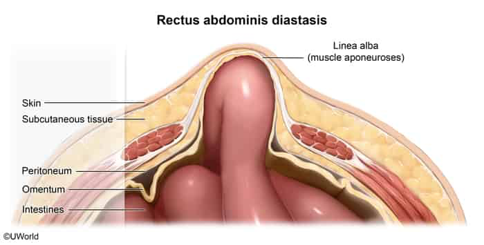
Widening and thinning of the linea alba with separation of the rectus abdominis muscles.
No true fascial defect is present (distinguishes it from a true hernia).
Risk Factors (RF) :
Multiparity / prior pregnancy (mechanical stretch + hormonal laxity).
Obesity / central adiposity.
Neonates / infants (normal physiologic finding, especially in premature infants).
Clinical Features :
Painless, longitudinal midline abdominal bulge (xiphoid to umbilicus).
Accentuates with ↑ intra-abdominal pressure (e.g., head raise / crunch maneuver , Valsalva, coughing).Flattens / disappears when the pt is relaxed and supine.
Diagnosis :
Clinical : Palpation of widened inter-rectus distance (> 2 cm) on physical exam.Imaging (US/CT) : Not routinely required; used only if diagnostic ambiguity exists to rule out a true hernia.
Key Differentials :
Umbilical / Ventral Hernia : Has a true fascial ring; carries risk of incarceration and strangulation .Rectus Sheath Hematoma : Acute, painful, unilateral; (+) Fothergill sign (mass remains fixed/palpable with abdominal wall contraction).
Management & High-Yield Pearls :
First-line : Reassurance and observation ± core-strengthening physical therapy.Neonates : Spontaneous resolution with abdominal wall development.Highest-Yield Exam Pearl : NO risk of bowel incarceration or strangulation because the linea alba remains intact (no true fascial defect) → not indicated.Refractory / Elective : Surgical plication / abdominoplasty (reserved strictly for severe cosmetic or functional impairment failing conservative therapy).