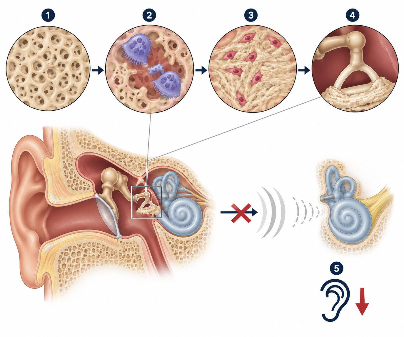
Most common cause of non-traumatic conductive hearing loss (CHL) in young/middle-aged adults (peak age 20–40 yo).
Female > Male (~2:1 ratio).
Autosomal dominant (AD) inheritance with incomplete penetrance (positive family history in ~50%).
Often accelerates during pregnancy or periods of high estrogen exposure.
Pathophysiology: Disordered bony remodeling (resorption followed by sclerosis) of the otic capsule -> progressive fixation of the stapes footplate to the oval window.
Clinical Features
Hearing Loss:
Progressive, insidious CHL (bilateral in 70–85%, though often asymmetric initially).
Predominantly affects low-frequency sounds early on.
Paracusis of Willisii: Paradoxical ability to hear speech better in noisy environments (high-yield buzzword).
Tinnitus: Low-frequency, buzzing, continuous (present in ~75%).
Vestibular symptoms: Mild dizziness or dysequilibrium (uncommon, ~10–20%).
Physical Exam:
Otoscopy: Usually normal tympanic membrane (TM).
Schwartze sign (“flamingo-pink” sign): Faint reddish/vascular hue over the promontory visible through the TM (indicates active, hyperemic phase of osteogenesis; seen in ~10%).
Tuning Fork Tests:
Rinne: Bone conduction > Air conduction (BC > AC) in the affected ear (confirms CHL).
Weber: Lateralizes to the affected ear (or to the worse ear if bilateral).
Diagnosis
Initial Test: Pure Tone Audiometry
Demonstrates air-bone gap (CHL).
Carhart notch: Characteristic drop in bone-conduction threshold at 2000 Hz (classic diagnostic finding).
Tympanometry:
Type As curve: Normal middle ear pressure with decreased peak compliance/amplitude due to ossicular chain stiffness.
Absent or elevated acoustic (stapedial) reflexes.
Confirmatory / Preoperative Imaging:
High-Resolution CT (HRCT) of temporal bone: Shows areas of demineralization/radiolucency in the otic capsule (most commonly at the fissula ante fenestram). Indicated for surgical planning or atypical presentations.
Differential Diagnostics
Presbycusis:
Diff: Elderly pts (>65 yo), sensorineural hearing loss (SNHL), high frequencies lost first, normal Rinne (AC > BC).
Cholesteatoma:
Diff: Otoscopy reveals retraction pocket with pearly-white keratin debris or granulation tissue; associated with chronic foul otorrhea.
Tympanosclerosis:
Diff: History of recurrent acute otitis media (AOM) or tympanostomy tubes; otoscopy shows chalky white, calcified plaques directly on the TM.
Ménière Disease:
Diff: Episodic vertigo (20 min to hours), fluctuating low-frequency SNHL (not CHL), and aural fullness.
Vestibular Schwannoma (Acoustic Neuroma):
Diff: Unilateral progressive SNHL, vertigo/imbalance, asymmetric sensorineural loss on audiometry, MRI shows cerebellopontine angle mass.
Management
First-Line / Conservative (Mild disease or non-surgical candidates):
Observation: For mild, unilateral, non-disruptive hearing loss.
Hearing aids (conventional amplification): Highly effective, non-invasive; preferred initial therapy if pt refuses surgery or high surgical risk.
Definitive / Surgical Management:
Stapedotomy or Stapedectomy: Fenestration or removal of the fixed stapes footplate with placement of a microprosthesis; treatment of choice for significant CHL with good speech discrimination.
Cochlear implantation: Indicated for profound mixed hearing loss or SNHL refractory to conventional amplification.
Note: Medical therapy (e.g., sodium fluoride, bisphosphonates) has limited efficacy and is generally not considered standard primary management on Step 2 CK.
Complications
Progressive mixed hearing loss or profound sensorineural hearing loss (“far-advanced otosclerosis” from cochlear invasion).
Surgical complications of stapedectomy:
Sensorineural deafness (rare, 1–2%).
Prolonged vertigo/balance disturbance.
Perilymph fistula.
Chorda tympani nerve injury -> metallic dysgeusia / loss of taste on anterior 2/3 of tongue.