Epidemiology & Risk Factors
↑ ICP without mass lesion, hydrocephalus, or abnormal CSF composition due to dysregulation of:
CSF production CSF absorption Cerebral venous outflow
Demographics : Young, obese females of reproductive age (F:M ratio ~9:1; BMI >30 kg/m²). c Medications (High-Yield Triggers) :
Tetracyclines (e.g., minocycline, doxycycline).Retinoids / Vitamin A toxicity (e.g., isotretinoin).Recombinant Growth Hormone (GH) .Excessive thyroid hormone replacement .Corticosteroid withdrawal .
Systemic Associations : SLE, CKD, hypoparathyroidism, sleep apnea.
Clinical Features
Headache : Holocranial, throbbing or dull, worse in the morning, exacerbated by Valsalva maneuvers (coughing, bending forward).Visual Symptoms :
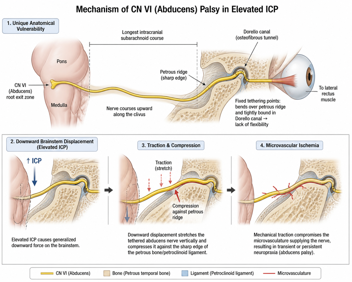
Transient visual obscurations (TVOs) : Brief episodes of visual dimming/blackouts lasting seconds, triggered by posture change.Diplopia : Binocular horizontal diplopia secondary to CN VI (abducens) palsy (false localizing sign due to traction from ↑ ICP). c
Auditory Symptoms : Pulsatile tinnitus (“whooshing” or wind-blowing sound synchronous with heartbeat).Physical Exam :
Bilateral papilledema (optic disc swelling w/ flame hemorrhages).
Visual field defects: Enlarged blind spot , peripheral visual field constriction.
Normal mental status, no other focal neurologic deficits.
Diagnosis
Step 1: Neuroimaging (Initial to rule out space-occupying lesion or hydrocephalus) :
Brain MRI with contrast + MRV : Essential before LP to rule out mass, herniation risk, or Cerebral Venous Sinus Thrombosis (CVST) .Typical neuroimaging findings of IIH: Empty sella turcica , flattening of posterior sclera/globes, distention of perioptic subarachnoid space, tortuous optic nerves.
Step 2: Lumbar Puncture (Confirmatory / Gold Standard) :
Performed in lateral decubitus position.
Opening pressure: >250 mm H₂O (or >25 cm H₂O; normal is <200 mm H₂O).CSF composition: Normal (normal cell count, protein, and glucose).
Monitoring :
Formal visual field testing (automated perimetry) : Required at baseline and serial follow-ups to detect insidious visual loss.
Differential Diagnostics
Cerebral Venous Sinus Thrombosis (CVST) : Diff by hypercoagulable risk factors (e.g., postpartum, OCP use), focal deficits, seizures; confirmed via MRV (flow void/filling defect in dural sinuses).Intracranial Neoplasm / Abscess : Diff by focal neuro signs, systemic symptoms (B-symptoms, fever), visible mass lesion with vasogenic edema on neuroimaging.Chronic Migraine : Diff by absence of papilledema, normal opening pressure on LP, photophobia/phonophobia, lack of pulsatile tinnitus.Giant Cell (Temporal) Arteritis : Diff by age >50, jaw claudication, scalp tenderness, markedly elevated ESR/CRP, temporal artery biopsy findings.Meningitis / Chronic Infection : Diff by fever, meningismus, abnormal CSF (pleocytosis, elevated protein, low glucose).
Management
First-Line Medical Therapy & Conservative Measures :
Weight Loss : 5–10% reduction of total body weight is the most effective long-term disease-modifying intervention.Acetazolamide (PO) : First-line pharmacotherapy (inhibits choroid plexus carbonic anhydrase → ↓ CSF production).Discontinue offending medications (stop isotretinoin, tetracyclines, etc.).
Second-Line / Adjunctive Therapy :
Furosemide : Added to acetazolamide if symptoms are refractory or acetazolamide is poorly tolerated.Short-course oral corticosteroids : Used solely as a temporizing bridge for acute, severe visual deterioration awaiting surgical decompression.Serial LPs : Not recommended for routine management (CSF regenerates within hours); used only for transient symptom relief in acute settings (e.g., during pregnancy).
Refractory Disease / Progressive Visual Loss (Surgical Interventions) :
Optic Nerve Sheath Fenestration (ONSF) : Indicated for rapidly progressive visual field defects refractory to maximum medical therapy.CSF Shunting (Ventriculoperitoneal [VP] or Lumboperitoneal [LP] shunt) : Indicated if severe, intractable headache persists or if visual loss worsens despite ONSF.Dural Venous Sinus Stenting : Emerging option if high-grade transverse sinus stenosis with elevated pressure gradient is present.
Complications
Permanent Visual Loss / Blindness : Most severe complication; caused by chronic optic nerve ischemia and subsequent optic atrophy.Drug-Induced Adverse Effects :
Acetazolamide : Paresthesias, hypokalemia, metabolic acidosis, renal calculi, nausea.
Shunt Complications : Catheter obstruction, infection/meningitis, low-pressure headaches.