Epidemiology & Risk Factors

  • Major cause of ischemic stroke in young/middle-aged patients (<45 yo).
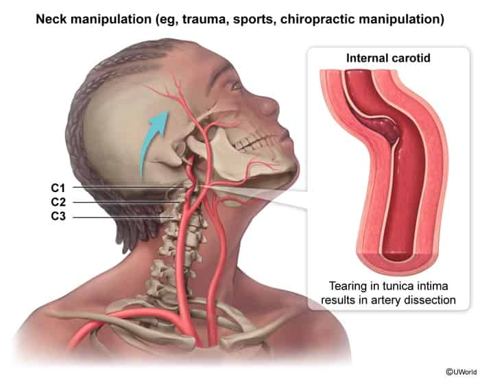
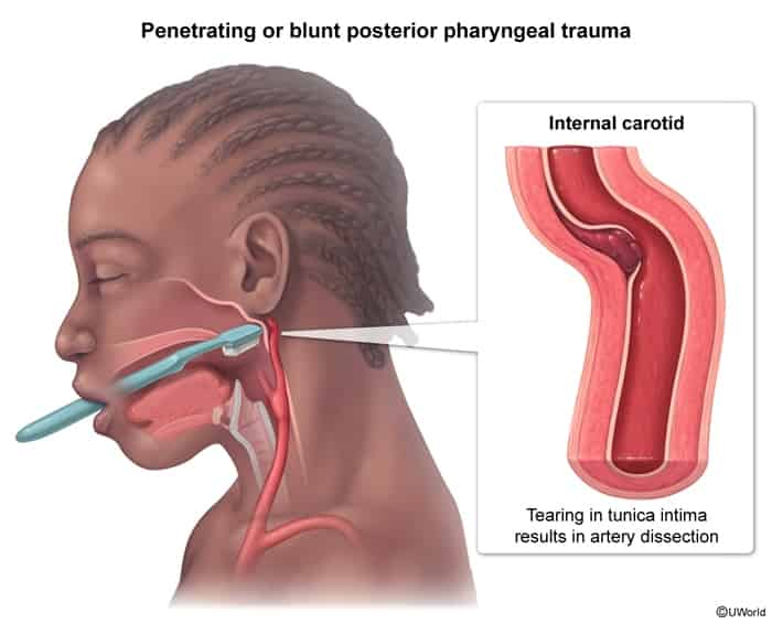
  • Mechanical/Trauma:
    • Carotid artery dissection, cervical spine manipulation (chiropractic), motor vehicle collisions (whiplash). c
    • Strenuous physical activity, yoga, coughing/vomiting paroxysms, rollercoasters.
  • Connective Tissue Disorders:
    • Fibromuscular Dysplasia (FMD) (high association).
    • Ehlers-Danlos syndrome (vascular type IV), Marfan syndrome, Osteogenesis Imperfecta.
  • Other: HTN, smoking, migraine history.

Clinical Features

  • Internal Carotid Artery Dissection (ICAD):
    • Unilateral frontotemporal/periorbital headache and ipsilateral neck pain.
    • Partial Horner syndrome: Ptosis + miosis without anhidrosis (facial sweat fibers travel along external carotid artery). c
    • Amaurosis fugax (transient monocular blindness) or retinal artery occlusion.
    • Pulsatile tinnitus, lower cranial nerve palsies (CN IX-XII, esp. CN XII / tongue deviation).
    • Ischemic signs: Contralateral motor/sensory deficits, aphasia (MCA/ACA territory).
  • Vertebral Artery Dissection (VAD):
    • Unilateral occipital headache and posterior neck pain.
    • Posterior circulation ischemia (Lateral Medullary / Wallenberg syndrome):
      • Ipsilateral ataxia, loss of facial pain/temperature sensation, Horner syndrome, dysphagia, hoarseness.
      • Contralateral body loss of pain/temperature sensation.
      • Vertigo, nystagmus, diplopia.

Diagnosis

  • Initial / Screening (Suspected acute stroke/dissection):
    • Non-contrast Head CT: Initial step in acute focal deficit to rule out hemorrhage before thrombolysis.
    • CTA of head and neck or MRA of head and neck: Initial diagnostic vascular imaging of choice (rapid, non-invasive, high sensitivity/specificity).
  • Confirmatory / Gold Standard:
    • Digital Subtraction Angiography (DSA) / Conventional catheter angiography (gold standard, but largely replaced by CTA/MRA).
  • Key Imaging Findings:
    • “String sign” (tapering long-segment stenosis).
    • “Flame sign” (tapered occlusion).
    • Crescentic intramural hematoma (seen best on axial T1 fat-saturated MRI).
    • Intimal flap, false lumen, or pseudoaneurysm.
  • Key Labs: CBC, PT/INR, PTT, BMP (renal function for CTA contrast), cardiac biomarkers.

Differential Diagnostics

  • Subarachnoid Hemorrhage (SAH):
    • Diff by: Sudden-onset “thunderclap” headache, meningismus, subarachnoid hyperdensity on non-contrast CT / RBCs on LP (xanthochromia).
  • Cluster Headache:
    • Diff by: Severe retro-orbital pain + autonomic signs (rhinorrhea, lacrimation), but transient/episodic (<3 hours) without focal neurological deficits or mural hematoma on imaging.
  • Giant Cell (Temporal) Arteritis:
    • Diff by: Age >50 yo, jaw claudication, scalp tenderness, markedly ↑ ESR/CRP, temporal artery biopsy.
  • Musculoskeletal Neck Strain:
    • Diff by: Absence of headache/Horner syndrome, normal vascular imaging, pain reproduced exclusively by mechanical palpation/movement.
  • Migraine with Aura:
    • Diff by: Gradual aura progression (<60 min) followed by pulsating headache with photophobia/nausea; lacks Horner syndrome or vascular imaging abnormalities.

Management

  • Acute Phase (<4.5 hours from ischemic stroke onset):
    • IV Alteplase (rtPA) or Tenecteplase: First-line for acute ischemic stroke within 4.5 hours if no contraindications (extracranial dissection is NOT a contraindication).
    • Endovascular Thrombectomy (EVT): Indicated for acute large vessel occlusion (LVO) of anterior circulation within eligible time windows (up to 24 hours).
  • Subacute / Maintenance Medical Therapy (First-line):
    • Antithrombotic Therapy for 3–6 months:
      • Antiplatelet therapy (e.g., Aspirin monotherapy or DAPT [Aspirin + Clopidogrel]) OR Anticoagulation (e.g., Heparin bridging to Warfarin/DOAC).
      • Note: Guidelines demonstrate equivalent stroke prevention efficacy between antiplatelet and anticoagulation. Antiplatelet is often preferred due to lower bleeding risk.
    • Strict BP control (avoid extreme hypertension to prevent hematoma extension; avoid excessive hypotension to maintain cerebral perfusion).
  • Refractory / Interventional (Second-line / Rescue):
    • Endovascular Angioplasty / Stenting: Indicated for:
      • Recurrent TIA/ischemic stroke despite optimal antithrombotic therapy.
      • Expanding dissecting pseudoaneurysm.
      • Hemodynamic compromise from severe luminal hypoperfusion.

Complications

  • Thromboembolic Ischemic Stroke / TIA (most common cause of long-term morbidity).
  • Dissecting Pseudoaneurysm (risk of distal embolization or local compressive neuropathy).
  • Subarachnoid Hemorrhage (SAH) (occurs if dissection extends intradurally beyond the dural ring).
  • Complete arterial occlusion leading to massive hemispheric or brainstem infarction.