Epidemiology
Etiology
CJD is caused by misfolded proteins (prions, PrPSc) that are either produced by affected individual themselves, or taken up from an exogenous source.
- Sporadic CJD (sCJD): Most common (~85%); typical onset 50–70 yo. c
- Familial CJD: Autosomal dominant mutation in the PRNP gene (~10–15%).
- Iatrogenic CJD: Contaminated surgical instruments, corneal grafts, dura mater grafts, or cadaveric human growth hormone.
- Variant CJD (vCJD): Associated with bovine spongiform encephalopathy (“mad cow disease”); presents in younger patients (<40 yo).
Pathophysiology
- Conversion of normal cellular prion proteins with alpha-helical structure (PrPc) to prions that demonstrate an increase in beta-pleated sheet structure (PrPSc) → conformational change of physiological PrPc → PrPSc accumulation and plaque formation → neuronal cell death → progression to spongiform encephalopathy
- Conformational change is triggered via misfolded PrPSc (from scrapie, a transmissible spongiform encephalopathy of sheep)
- Since misfolded prions are insoluble, they deposit as plaques resistant to proteases and standard autoclaving, thus contributing to the formation of more PrPSc.
Clinical features
- Rapidly progressive dementia: Cognitive decline progressing over weeks to months (hallmark).
- Myoclonus: Involuntary muscle jerks, characteristically startle-induced by auditory or tactile stimuli. c
- Cerebellar & Extrapyramidal dysfunction:
- Ataxia, dysmetria, and gait instability (often early).
- Rigidity, bradykinesia, tremor, choreoathetosis.
- Neuropsychiatric symptoms: Early mood changes, apathy, anxiety, psychosis, or visual hallucinations.
- Akinetic mutism: Pt becomes unresponsive, immobile, and mute (late stage).
- Visual disturbances: Cortical blindness, diplopia, or visual field deficits (Heidenhain variant).
Tip
Rapidly progressive dementia and myoclonic jerks are the hallmarks of Creutzfeldt-Jakob disease.
Diagnostics
- Initial / Best Initial Imaging: Brain MRI (DWI / FLAIR)
- “Cortical ribboning” (hyperintensity of cerebral cortex). c
- Hyperintensity in basal ganglia (putamen and caudate head).
- “Pulvinar sign” or “hockey-stick sign” in posterior thalamus (more common in vCJD).
- Lumbar Puncture (LP) & CSF Analysis:
- Real-time quaking-induced conversion (RT-QuIC): Most sensitive and specific CSF diagnostic test.
- 14-3-3 protein: Elevated (supportive; non-specific marker of rapid neuronal lysis).
- Total tau protein: Markedly elevated.
- Standard CSF (WBC, glucose, total protein) usually unremarkable/normal.
- Electroencephalogram (EEG):
- Periodic sharp wave complexes (PSWCs) (synchronous, biphasic/triphasic discharges at 1–2 Hz).
- Confirmatory / Gold Standard:
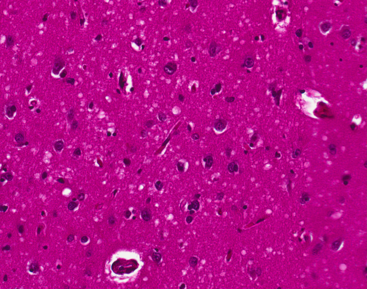
- Brain biopsy / Post-mortem autopsy: Shows spongiform degeneration/vacuolization of neuropil and astrocytic gliosis without inflammatory infiltration.
- Note: Biopsy rarely performed premortem due to diagnostic accuracy of non-invasive tests and transmission risks to healthcare staff.
Differential Diagnostics
- Alzheimer Disease:
- Differentiated by insidious onset and slow progression over years (vs weeks to months in CJD); no startle myoclonus; normal RT-QuIC and MRI diffusion.
- Autoimmune / Paraneoplastic Encephalitis (e.g., Anti-NMDA receptor):
- Differentiated by CSF pleocytosis/oligoclonal bands, presence of specific autoantibodies, and improvement with immunotherapy.
- Hashimoto Encephalopathy (SREAT):
- Differentiated by marked elevation of anti-TPO antibodies and dramatic clinical response to high-dose IV corticosteroids.
- Normal Pressure Hydrocephalus (NPH):
- Differentiated by triad of “wet, wacky, wobbly” (gait ataxia, urinary incontinence, cognitive decline), ventriculomegaly out of proportion to sulcal enlargement on imaging, and positive response to high-volume LP.
- Infectious Encephalitis (e.g., HSV, HIV, PML):
- Differentiated by fever, systemic signs, CSF pleocytosis, positive PCR (e.g., HSV DNA), or focal temporal lobe lesions (HSV) vs JC virus demyelination (PML).
Treatment
- Primary / First-Line:
- Palliative & Supportive Care: No curative or disease-modifying therapy exists; median survival is 6–12 months from symptom onset.
- Early goals-of-care discussions, advance directives, and hospice referral.
- Symptom Control:
- Myoclonus: Clonazepam (first-line) or Levetiracetam / Valproic acid.
- Agitation/Psychosis: Low-dose atypical antipsychotics (e.g., Quetiapine) with caution.
- Infection Control Precautions:
- Standard sterilization (autoclaving/alcohol) is insufficient; requires specialized protocols (e.g., sodium hydroxide immersion + high-temperature autoclaving) for neurosurgical instruments to prevent iatrogenic spread.