Open globe injury
- Etiology & Risk Factors:
- High-velocity projectiles (e.g., hammering metal-on-metal, grinding), penetrating/blunt trauma, falls.
- Lack of eye protection; prior ocular surgery (structural weak point).
- Clinical Features:
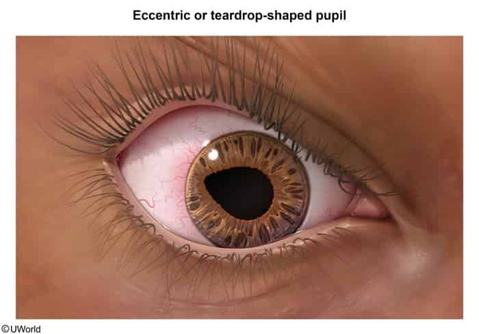
- Teardrop/peaked pupil (eccentric pupil pointing toward the wound).
- Extrusion of intraocular contents (uveal tissue, iris prolapse, vitreous leakage).
- Shallow or flattened anterior chamber (or deep if posterior scleral rupture).
- Positive Seidel test (fluorescein streaming/dilution from aqueous leak; do not perform if overt).
- 360° bullous subconjunctival hemorrhage, hyphema, acute loss of visual acuity.
- Diagnostic Workup & Contraindications:
- Best imaging: Non-contrast thin-cut CT of the orbits (detects occult rupture and radiopaque intraocular foreign bodies [IOFB]).
- CONTRAINDICATED:
- No tonometry (measuring IOP triggers intraocular extrusion).
- No eye manipulation (do not retract eyelids or place direct pressure).
- No MRI (risk of projectile movement if metallic IOFB is present).
- No patches or gauze placed directly onto the globe.
- Acute Management:
- Rigid eye shield (e.g., Fox shield) resting strictly on orbital bony margins.
- NPO for urgent surgical exploration (<24 hours).
- Elevate HOB 30–45° to minimize intraocular pressure.
- Antiemetics (IV ondansetron) and analgesia to prevent Valsalva-induced extrusion from vomiting/coughing/crying.
- IV broad-spectrum antibiotics (e.g., Vancomycin + Ceftazidime) for endophthalmitis prophylaxis.
- Tetanus toxoid booster ± TIG.
- Emergent ophthalmology consult for definitive primary surgical closure.
- High-Yield Complications:
- Endophthalmitis: Severe bacterial/fungal intraocular infection (Bacillus cereus, Staphylococcus).
- Sympathetic ophthalmia: Autoimmune bilateral granulomatous panuveitis caused by immune sensitization to sequestered uveal/retinal antigens exposed during trauma; damages the contralateral uninjured eye weeks to months later.
- Retinal detachment and permanent visual loss.