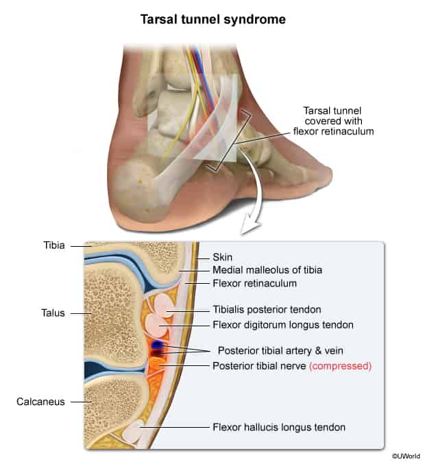
Pathophysiology: Entrapment/compression neuropathy of the tibial nerve (or its terminal branches: medial and lateral plantar nerves) within the fibro-osseous tarsal tunnel posterior/inferior to the medial malleolus.
Burning pain, paresthesias, and numbness over the plantar aspect of the foot, heel, and toes.
Symptoms typically worsen with prolonged weight-bearing, walking, or at night.
Proximal radiation of pain up the posteromedial calf (Valleix phenomenon).
Physical Examination:
(+) Tinel sign: Reproduction of radiating paresthesias to the sole/toes upon percussion of the tibial nerve posterior/inferior to the medial malleolus.
Pain exacerbated by forced passive dorsiflexion and eversion (stretches the tibial nerve) or sustained plantar flexion-inversion. c
Sensory loss restricted to the plantar foot (dorsum of foot is spared; innervated by peroneal nerve).
Advanced/chronic disease: Atrophy and weakness of intrinsic foot muscles (e.g., weak toe flexion, clawing of toes).
Diagnosis
Initial/Clinical: Primarily a clinical diagnosis based on characteristic Hx, plantar sensory symptoms, and a (+) Tinel sign at the medial malleolus.
Confirmatory / Gold Standard: Electromyography & Nerve Conduction Studies (EMG/NCS) (demonstrates prolonged motor/sensory latencies or decreased conduction velocity/amplitudes of the medial/lateral plantar nerves).
Imaging:
Plain Radiographs (X-ray ankle/foot): Initial imaging to rule out bony pathology, fractures, osteophytes, or structural deformities.
MRI / High-Resolution US of the ankle: Best modalities to identify soft tissue etiologies (ganglion cyst, schwannoma, tendon sheath effusion, flexor retinaculum thickening).
Differential Diagnostics
Plantar Fasciitis:
Diff: Pain localized strictly to the plantar/medial calcaneal insertion, maximal with the first steps in the morning or after periods of rest; lacks numbness/paresthesias; (-) Tinel sign at tarsal tunnel.
Morton Neuroma:
Diff: Pain and numbness localized between the 3rd and 4th metatarsal heads (interdigital); reproduced by transverse foot compression with a palpable/audible click (Mulder sign); hindfoot examination normal.
L5–S1 Radiculopathy (Sciatica):
Diff: Back pain radiating down the posterior/lateral leg; (+) Straight Leg Raise (SLR); sensory and motor deficits extend above the ankle into the leg/thigh; diminished/absent Achilles reflex (S1).
Diabetic Peripheral Neuropathy:
Diff: Bilateral, symmetric, “stocking-glove” sensory loss; not confined to a single peripheral nerve distribution; no focal tenderness/Tinel sign at the medial malleolus.
Achilles Tendinopathy:
Diff: Posterior heel pain aggravated by push-off or resisted plantarflexion; tenderness over the Achilles tendon substance or insertion; no neurosensory deficits.
Management
First-Line (Conservative):
Activity modification, rest, and NSAIDs for pain/inflammation control.
Orthotics: Medial arch supports (to correct pes planus/eversion) and semi-rigid foot orthoses to offload the medial ankle.
Night splints or a walking boot for immobilization.
Local corticosteroid injection into the tarsal tunnel (guided by ultrasound; avoids direct intraneural injection) for persistent pain/inflammation.
Refractory / Surgical:
Surgical decompression (Tarsal Tunnel Release): Flexor retinaculum division ± release of the deep fascia and neurolysis of the medial/lateral plantar and calcaneal branches.
Indicated if: Failure of conservative management (typically > 3–6 months), progressive motor/sensory deficits, or identifiable compressive mass lesion (e.g., cyst excision).
Complications
Permanent neuropathic pain and sensory loss over the plantar surface.
Intrinsic foot muscle atrophy and contractures (claw toe deformities).
Chronic Regional Pain Syndrome (CRPS) of the foot/ankle.
Postoperative recurrence secondary to perineural fibrosis and scar tissue tethering.