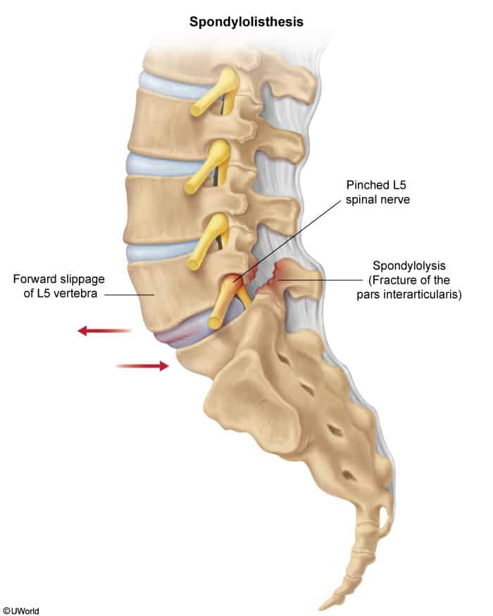
Definition: Anterior (or posterior) displacement of a superior vertebral body relative to the inferior vertebra (most commonly L5-S1 in younger pts, L4-L5 in older pts).
Types & Risk Factors:
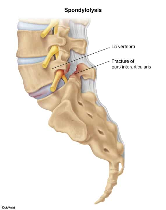
Isthmic: Due to bilateral spondylolysis (stress fracture of pars interarticularis). Common in adolescent athletes with repetitive lumbar hyperextension (e.g., gymnastics, diving, football linemen, figure skating).
Degenerative: Secondary to facet joint osteoarthritis (OA) and disc degeneration. Most common in females > 50 yo at L4-L5.
Dysplastic (Congenital): Congenital malformation of lumbosacral junction.
Traumatic: High-energy acute fracture of posterior elements.
Pathologic: Malignancy, infection, or metabolic bone disease (e.g., Paget disease, osteogenesis imperfecta).
Clinical Features
History:
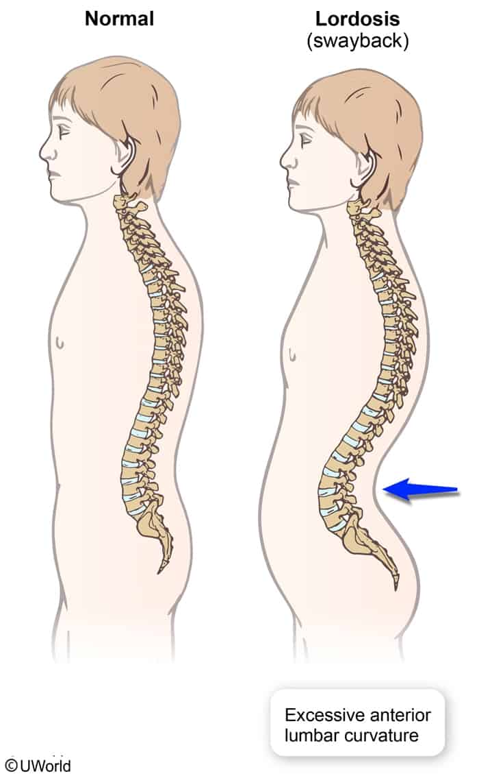
Insidious-onset chronic lower back pain (LBP) aggravated by lumbar extension, standing, and walking; relieved by lumbar flexion and sitting/rest. c
Radiculopathy: Sciatica, buttock/thigh pain, paresthesias, or neurogenic claudication (exiting L5 or traversing S1 nerve root compression).
Physical Exam:
Palpable “step-off” deformity over the lumbar spine at the level of the slip (in moderate-to-severe slips).
Hamstring tightness/spasm leading to a crouched gait (Phalen-Dickson gait).
Lumbar tenderness elicited with hyperextension (positive stork test / single-leg hyperextension).
Focal neurologic deficits: Motor weakness (e.g., great toe dorsiflexion weakness for L5), sensory loss, or diminished deep tendon reflexes (DTRs, e.g., Achilles reflex for S1).
Diagnosis
Initial/Screening:
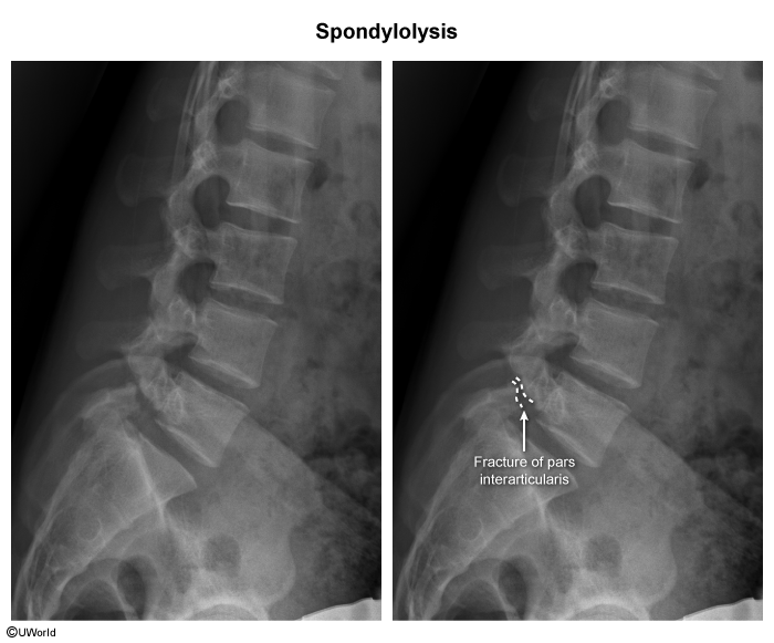
Standing Plain Radiography (XR) of Lumbar Spine: AP, lateral, and dynamic flexion-extension views. c
Lateral XR establishes diagnosis and grading via Meyerding Classification:
Grade I: 0–25% slip.
Grade II: 26–50% slip.
Grade III: 51–75% slip.
Grade IV: 76–100% slip.
Grade V: > 100% (spondyloptosis).
Oblique XR: May show the classic “collar on the Scottie dog” sign (indicates pars interarticularis fracture/spondylolysis).
Confirmatory / Advanced Imaging:
Lumbar Spine MRI: Gold standard for evaluating spinal cord/cauda equina compression, central or neuroforaminal canal stenosis, disc herniation, and soft tissue pathology. Indicated if radicular symptoms or neurologic deficits are present.
CT Lumbar Spine: Most sensitive for fine bony detail, non-union of pars fracture, or when MRI is contraindicated.
Key Labs:
Typically normal (CBC, ESR, CRP used only to rule out osteomyelitis/epidural abscess or malignancy).
Differential Diagnostics
Spondylolysis:
Bony defect in the pars interarticulariswithout slippage/displacement of the vertebral body. No palpable step-off.
Lumbar Spinal Stenosis:
Neurogenic claudication relieved by flexion (“shopping cart sign”), but typically lacks a palpable structural step-off on PE unless degenerative spondylolisthesis is also present.
Lumbar Disc Herniation:
Pain is worsened by lumbar flexion and sitting (vs extension in spondylolisthesis); positive Straight Leg Raise (SLR) test.
Ankylosing Spondylitis:
Morning stiffness lasting > 30 min, improves with exercise/activity, young adult males, (+) HLA-B27, sacroiliitis on pelvic XR.
Cauda Equina Syndrome:
Severe surgical emergency; presents with saddle anesthesia, acute urinary retention/overflow incontinence, fecal incontinence, and bilateral lower extremity flaccid paralysis.
Epidural Steroid Injections (ESI) or facet joint injections for targeted neuropathic pain relief and inflammation reduction.
Refractory / Surgical Management:
Indications:
High-grade slip (Grade III, IV, or V).
Progressive or severe neurologic deficits (e.g., motor weakness, foot drop).
Cauda equina syndrome (Emergent).
Intractable pain failing ≥ 6 months of conservative therapy.
Procedure: Decompressive laminectomy with instrumented spinal fusion (posterolateral or interbody fusion, e.g., TLIF/PLIF) with or without slip reduction.
Complications
Permanent neurological deficit (e.g., chronic radiculopathy, motor paresis, foot drop).
Cauda Equina Syndrome (central canal compromise from high-grade slip).
Progression to Spondyloptosis (complete displacement of L5 anterior to S1).
Failed back surgery syndrome or adjacent segment disease post-fusion.