Seborrheic keratosis is a benign growth of immature keratinocytes. The name is essentially a historical misnomer; Although the lesions of seborrheic keratosis appear greasy, they are not caused by sebum.
Epidemiology
Seborrheic keratosis is the most common benign skin tumor in adults > 30 years of age.
Etiology
- Associated with activating mutations of the fibroblast growth factor receptor 3.
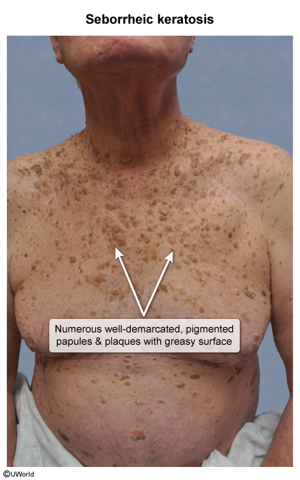
- SK itself is benign, but rapid onset of numerous SKs (Leser-Trélat sign) is often associated with an internal malignancy (eg, gastric adenocarcinoma), possibly due to overproduction of insulin-like growth factor 1 and other cytokines.
Tip
Don’t mix with Mucocutaneous lentigines in Peutz-Jeghers syndrome
Pathophysiology
Clinical features
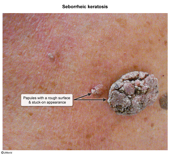
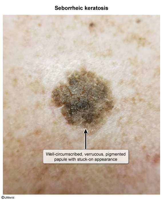
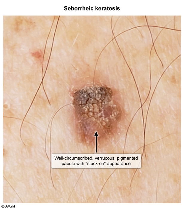
- Appearance: Well-circumscribed, round or oval macules, papules, or plaques.
- Classic Description: Dull, waxy, verrucous, velvety, or crumbly surface with a distinct “stuck-on” appearance. c
- Color: Highly variable; flesh-colored, tan, brown, to deeply pigmented/black.
- Distribution: Trunk, face, neck, and extremities (spares palms and soles).
- Symptoms: Typically asymptomatic; can become irritated, pruritic, or bleed with minor friction/trauma.
Diagnostics
- Initial/Screening: Clinical diagnosis based on gross visual inspection and dermoscopy.
- Dermoscopy findings: Milia-like cysts, comedo-like openings, and “brain-like” (cerebriform) pattern.
- Confirmatory/Gold Standard: Excisional or shave biopsy (only indicated if lesion is atypical, changing, bleeding, or cannot be distinguished from melanoma).
- Histology: Hyperkeratosis, papillomatosis, acanthosis, and intraepidermal pseudo-horn cysts (keratin-filled invaginations).
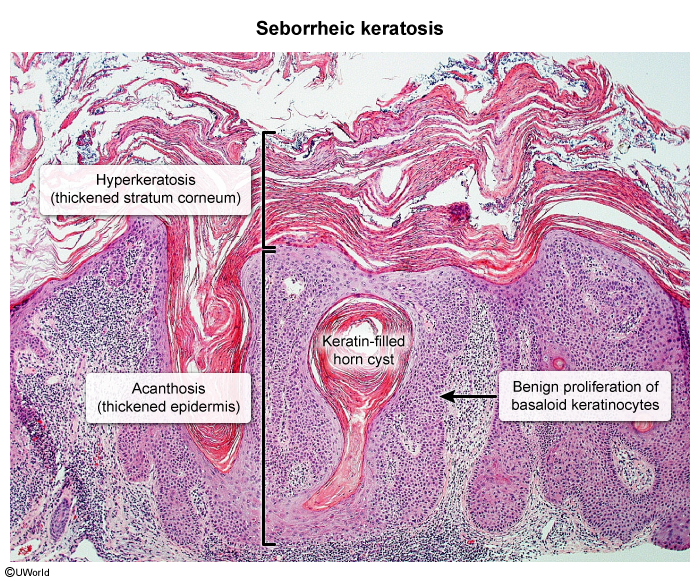
- Histology: Hyperkeratosis, papillomatosis, acanthosis, and intraepidermal pseudo-horn cysts (keratin-filled invaginations).
- Key Labs/Imaging:
- None required for isolated, typical SKs.
- If suspected Leser-Trélat sign: Age-appropriate cancer screening, GI endoscopy (EGD/colonoscopy), and CT chest/abdomen/pelvis.
Treatment
- First-line: Observation & reassurance (benign lesion with no malignant potential).
- Second-line (Symptomatic / Cosmesis / Patient preference):
- Cryotherapy (liquid nitrogen spray): Fast, effective, standard office procedure.
- Curettage / Shave excision / Electrodessication: Useful when histopathologic confirmation is desired.
- Topical hydrogen peroxide 40% solution (FDA-approved, alternative non-surgical option).
- Refractory / Suspicious Lesions:
- Complete shave or punch biopsy to rule out malignancy.
- Leser-Trélat Presentation:
- Treat the primary underlying internal malignancy (SKs often regress following resection/systemic therapy).