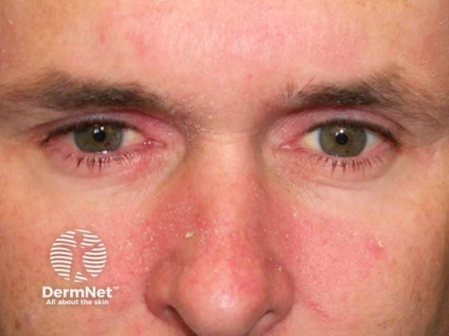
Bimodal distribution: Infancy (first 3 mos, self-limiting) and Adulthood (30-60 yo; predilection for males).
Associated conditions (High-Yield NBME triggers):
Parkinson disease and other neuro conditions (e.g., stroke, TBI).
HIV infection (severe, sudden-onset, widespread, or refractory presentation warrants HIV screening).
Pathophysiology: Inflammatory response to commensal Malassezia species (lipophilic yeast) in areas of high sebaceous gland density; non-contagious. c
Clinical Features
Infants:
“Cradle cap”: Non-pruritic, asymptomatic, thick, greasy, yellowish scales over the scalp vertex.
May involve diaper area, face, and intertriginous neck folds (erythema with greasy maceration; typically spares groin creases).
Adults:
Erythematous patches/plaques with greasy, yellowish, oily scales or fine, white, dry flaking (dandruff/pityriasis capitis).
Distribution: Sebum-rich areas:
Scalp, hairline, and external auditory canal.
Nasolabial folds, glabella, eyebrows, and beard area.
Presternal area and upper back.
Symptoms: Mild-to-moderate pruritus, burning, worsened during cold/dry weather or periods of emotional/physical stress.
Diagnosis
Initial/Screening: Clinical diagnosis based on characteristic appearance and anatomical distribution.
Key Labs:
None routinely indicated.
HIV 4th-gen Ag/Ab combo assay: Indicated if lesions are atypical, explosive in onset, severe, or treatment-refractory.
Confirmatory/Gold Standard: Clinical; skin biopsy rarely required (shows hyperkeratosis, parakeratosis around follicles, and spongiosis).
Ancillary:
KOH preparation: Performed to rule out dermatophyte infections (e.g., tinea); negative for branching pseudohyphae/hyphae characteristic of dermatophytosis.
Differential Diagnostics
Psoriasis:
Diff by well-demarcated plaques with thick, silvery-white (micaceous) scales, Auspitz sign (pinpoint bleeding upon scraping scale), extensor distribution (elbows, knees), and nail pitting.
Atopic Dermatitis:
Diff by severe, intractable pruritus, predilection for flexural creases (antecubital/popliteal fossae in adults/older kids) vs cheeks/extensors in infants (spares diaper area), and strong personal/atopic family history.
Tinea Capitis:
Diff by localized scaly patches with alopecia, “black dots” (broken hair shafts), and cervical/postauricular lymphadenopathy; (+) KOH mount for branching fungal hyphae.
Rosacea:
Diff by persistent central facial erythema, flushing provoked by hot liquids/spicy food/alcohol, telangiectasias, and papulopustules without comedones or greasy scaling.
Langerhans Cell Histiocytosis:
Diff in infants by refractory, weeping, purpuric/petechial scalp and diaper rash associated with systemic signs (lytic bone lesions, hepatosplenomegaly, failure to thrive).
Management
Infants (“Cradle Cap”):
First-line (Conservative): Emollients (mineral oil, petroleum jelly, baby oil) to soften crusts, followed by gentle massage with a soft brush and non-medicated baby shampoo.
Second-line/Refractory: Short course of low-potency topical hydrocortisone 1% or ketoconazole 2% cream/shampoo.
Adults (Scalp):
First-line: Topical antifungals: Ketoconazole 2% shampoo, selenium sulfide, zinc pyrithione, or tar-based shampoos (used 2-3x/week). c
Second-line/Acute Flare: Intermittent, short-term low-to-medium potency topical corticosteroids (e.g., fluocinolone, betamethasone lotion) to control pruritus and erythema.