Epidemiology & Risk Factors
- Most common inherited retinal dystrophy; onset in childhood to young adulthood.
- Progressive hereditary dystrophy of the retina or of the photoreceptors and the retinal pigment epithelium
- Inheritance: AD, AR, or XL (most severe).
- Syndromic associations:
- Usher syndrome: RP + congenital sensorineural hearing loss.
- Bardet-Biedl syndrome: RP + obesity, polydactyly, hypogonadism, renal anomalies.
Clinical Features
- Symptoms:
- Nyctalopia (night blindness): Earliest presentation (rod degeneration). c
- Progressive visual field loss: Concentric peripheral constriction (“tunnel vision”). Central vision lost late.
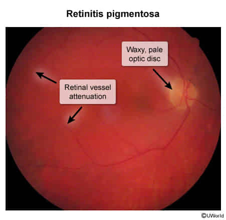
- Fundoscopic Triad: c
- “Bone-spicule” pigment deposits (mid-periphery).
- Attenuated retinal arterioles.
- Waxy pallor of the optic disc.
Diagnosis
- Initial: Dilated fundoscopy (classic triad) + Visual field perimetry (symmetric ring scotoma/constriction).
- Confirmatory / Gold Standard: Electroretinography (ERG) (reduced a- and b-wave amplitudes; rods extinguished before cones).
- Adjunctive:
- OCT: Assesses photoreceptor loss and detects cystoid macular edema (CME).
- Genetic testing: Evaluates for targetable mutations (e.g., RPE65).
Differential Diagnostics
- Open-Angle Glaucoma: Diff by optic disc cupping (↑ cup-to-disc ratio), elevated IOP, absence of bone spicules.
- Hydroxychloroquine Retinopathy: Diff by drug exposure history, “bull’s-eye” maculopathy, primarily central/paracentral defects.
- Vitamin A Deficiency: Diff by Bitot spots (conjunctival plaques), xerophthalmia; reversible with repletion. c
Management
- Targeted: Voretigene neparvovec (gene therapy) for confirmed biallelic RPE65 mutations.
- Supportive: UV-blocking sunglasses, low-vision aids, mobility training, genetic counseling.
- Secondary Complications:
- CME: Carbonic anhydrase inhibitors (oral acetazolamide or topical dorzolamide).
- Cataracts: Surgical lens extraction with IOL placement.
Complications
- Cystoid macular edema (CME) (leading treatable cause of acute visual acuity decline).
- Posterior subcapsular cataracts.
- Legal blindness (visual field ≤ 20° or visual acuity ≤ 20/200).