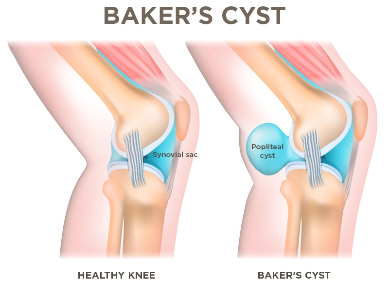
Adults: Secondary to intra-articular knee pathology (excessive synovial fluid production with valve-like flow into gastrocnemius-semimembranosus bursa).
Osteoarthritis (OA) (most common).
Meniscal tears (especially medial meniscus).
Rheumatoid arthritis (RA) and other inflammatory arthritides.
Prior knee trauma or ligamentous injury (ACL).
Pediatrics: Often primary/idiopathic; typically benign, asymptomatic, and self-limiting without underlying joint pathology.
Clinical Features
History:
Posterior knee fullness, tightness, or dull ache.
Symptoms worsen with prolonged standing, walking, or full knee extension.
Transient fluctuations in cyst size.
Physical Examination:
Palpable, fluctuant, non-pulsatile fullness at the posteromedial popliteal fossa.
Foucher sign: Mass becomes firm/tense with knee full extension and softer/less prominent with knee flexion (45°).
Sudden onset severe posterior knee and calf pain with erythema, warmth, and edema (mimics acute DVT).
Crescent sign: Ecchymosis distal to the medial or lateral malleolus caused by tracking of synovial fluid/blood.
Diagnosis
Initial / Best Imaging: Duplex Ultrasonography (US).
Demonstrates anechoic/hypoechoic fluid-filled cystic mass with a neck communicating with the joint space between the semimembranosus tendon and medial head of the gastrocnemius.
Crucial to exclude Deep Vein Thrombosis (DVT), particularly when ruptured.
Arthrocentesis / Cyst aspiration: Not routinely required; indicated if septic arthritis or infection is suspected (leukocyte count, Gram stain, culture).
Inflammatory markers (ESR/CRP): Elevated in underlying RA, crystal arthropathy, or infection.
Differential Diagnostics
Deep Vein Thrombosis (DVT):
Differentiating features: Non-compressible popliteal/femoral vein on Doppler US, absence of discrete cystic mass, (+) D-dimer. Ruptured cyst clinically mimics DVT completely; US is mandatory to distinguish.
Popliteal Artery Aneurysm:
Differentiating features: Pulsatile mass with arterial thrill/bruit, bilateral in ~50% of cases, arterial flow on color Doppler US.
Gastrocnemius / Plantaris Tear:
Differentiating features: Acute onset “pop” during explosive exertion (e.g., tennis); hematoma and disrupted muscle fibers on US/MRI without intra-articular communication.
Soft Tissue Sarcoma / Synovial Sarcoma:
Differentiating features: Firm, fixed, non-tender, non-transilluminating solid mass; does not change size with knee flexion/extension; solid tissue enhancement on MRI.
Ganglion Cyst:
Differentiating features: Lacks communication with the knee joint space; can arise from tendon sheaths or cruciate ligaments.
Management
Asymptomatic:
Reassurance and observation (spontaneous resolution common, especially in children).
Symptomatic (First-line / Conservative):
RICE (Rest, Ice, Compression, Elevation) and activity modification.
NSAIDs for pain and local inflammation.
Physical therapy focusing on hamstring/quadriceps flexibility and ROM.
Targeted therapy for underlying cause (e.g., OA or RA optimization).