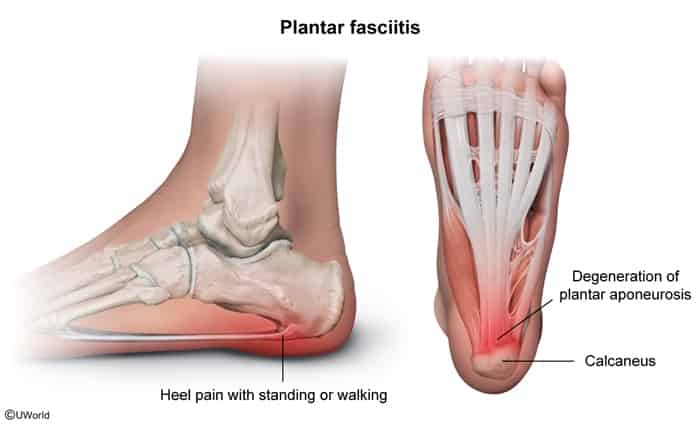
Worst with the first few steps in the morning or after prolonged periods of inactivity/rest.
Pain typically lessens with moderate activity/walking, but worsens toward the end of the day or after prolonged weight-bearing.
Physical Examination (PE):
Point tenderness to palpation over the anteromedial calcaneus at the plantar fascia insertion.
Pain exacerbated by passive dorsiflexion of the toes / great toe (Windlass test).
Plantar fascia may feel indurated or tense.
Diagnosis
Initial / Screening:
Clinical diagnosis based on classic Hx and PE (no routine imaging required).
Imaging:
Foot X-ray (Weight-bearing):
Indicated if red flags present, atypical features, or persistent pain despite conservative therapy to r/o bone pathology (e.g., stress fx).
May show a calcaneal spur (heel spur); incidental finding and not the cause of pain. c
Ultrasound / MRI Foot:
Reserved for refractory cases (> 3-6 mo) or suspected fascia rupture.
Shows thickening of the plantar fascia (> 4 mm) and perifascial fluid/edema.
Key Labs:
Routine labs are normal (negative inflammatory markers).
Differential Diagnostics
Calcaneal Stress Fracture:
Diff by pain elicited with the calcaneal squeeze test (mediolateral compression of the heel), pain worsening throughout the entire duration of weight-bearing (not just first steps), and focal bony tenderness.
Diff by burning pain, numbness, and paresthesias radiating to the plantar sole; (+) Tinel sign posterior to the medial malleolus; pain often worse at night.
Achilles Tendinopathy / Enthesitis:
Diff by pain and tenderness localized to the posterior heel (Achilles insertion) rather than the inferior/plantar aspect; associated with seronegative spondyloarthropathies (e.g., Ankylosing Spondylitis, Reactive Arthritis).
Heel Fat Pad Atrophy:
Diff by deep, diffuse aching directly under the central heel (not medial tubercle); worse when walking barefoot on hard surfaces; pain increases progressively with standing without a morning peak.
Plantar Fascia Rupture:
Diff by sudden “pop” or acute onset of severe pain, often following a corticosteroid injection, accompanied by localized hematoma and palpable fascia defect.
Management
First-line (Conservative Therapy):
Plantar fascia-specific stretching and calf/Achilles tendon stretching exercises.