Pediatric Fractures

Epidemiology & Risk Factors

  • Anatomic differences in pediatric bone:
    • Thick, highly vascular periosteum (limits displacement, promotes rapid remodeling).
    • High collagen-to-mineral ratio (more pliable, leads to incomplete/plastic deformation).
    • Presence of physis (growth plate) (weakest point of pediatric musculoskeletal system; vulnerable to shear stress).
  • Common Etiologies:
    • Accidental: Falls on outstretched hand (FOOSH), sports, playground injuries, birth trauma (clavicle).
    • Non-accidental trauma (NAT / Child Abuse): Age < 1-2 yo, non-ambulatory infants, inconsistent history, delayed presentation.

Clinical Features

  • Supracondylar Humeral Fracture:
    • Most common pediatric elbow fracture (peak age: 5-8 yo).
    • Mechanism: FOOSH with hyperextended elbow.
    • Exam: Gross elbow deformity, prominent swelling, ecchymosis over anterior cubital fossa.
    • Neurovascular risks:
      • Brachial artery injury (absent or diminished radial pulse).
      • Median nerve / Anterior Interosseous Nerve (AIN) injury (most common; loss of “OK” sign / motor weakness of flexor pollicis longus and flexor digitorum profundus).
      • Radial nerve injury (wrist drop, loss of dorsal hand sensation).
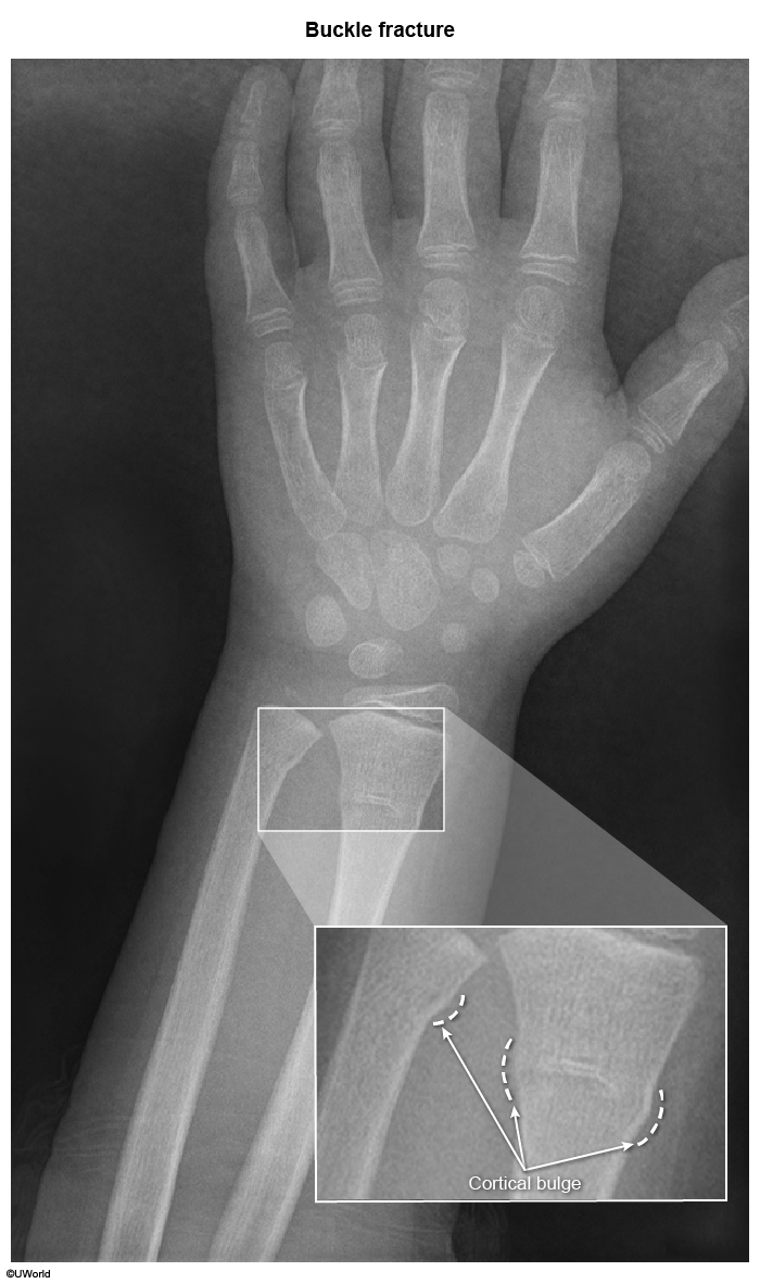
  • Torus (Buckle) Fracture:
    • Mechanism: Low-energy axial loading (FOOSH).
    • Exam: Mild focal tenderness, intact ROM, no gross deformity.
    • Pathology: Compression of metaphysis causing cortical bulging without true cortical breach.
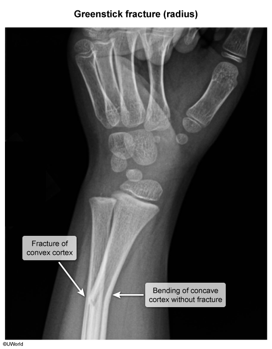
  • Greenstick Fracture:
    • Mechanism: Bending force applied to bone.
    • Pathology: Incomplete fracture with cortical breach on the convex (tension) side; intact cortex and periosteum on the concave (compression) side.
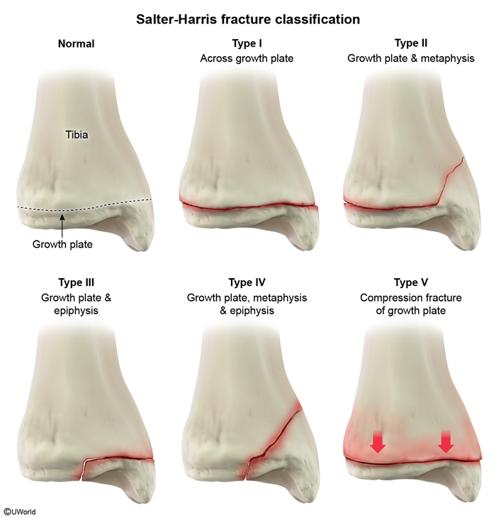
  • Physeal Fractures (Salter-Harris Classification):
    • Type I (Slipped): Physis separation only; normal or subtle widening on X-ray; excellent prognosis.
    • Type II (Above): Physis + Metaphysis (most common, ~75%); good prognosis.
    • Type III (Lower): Physis + Epiphysis (intra-articular); requires anatomical reduction.
    • Type IV (Through): Metaphysis + Physis + Epiphysis (intra-articular); high risk for premature physeal closure.
    • Type V (cRushed): Axial compression/crush of physis; worst prognosis (growth arrest / limb discrepancy).
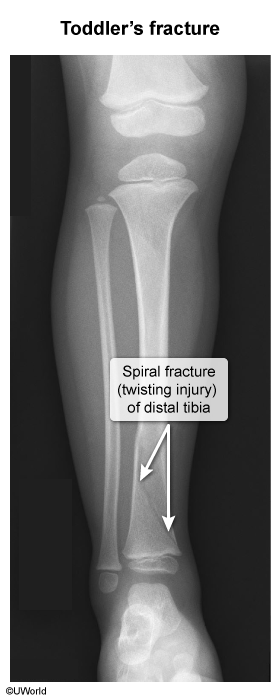
  • Toddler’s Fracture (Tibial Spiral Fracture):
    • Age: 9 months – 3 years (newly ambulatory).
    • Mechanism: Low-energy twisting fall while walking/running.
    • Exam: Refusal to bear weight, limp, mild localized tibial warmth/tenderness; absent gross deformity.
  • Clavicular Fracture:
    • Mechanism: Birth trauma (macrosomia, shoulder dystocia) or direct fall on shoulder.
    • Exam: Decreased active movement of arm (pseudoparalysis), crepitus, asymmetric Moro reflex (intact biceps/grasp reflex distinguishes from brachial plexus palsy).
  • Fractures Suspicious for NAT / Abuse:
    • Posterior rib fractures (pathognomonic; squeeze mechanism).
    • Metaphyseal corner / “bucket-handle” fractures (torsion/traction from pulling or shaking).
    • Multiple fractures in different stages of healing.
    • Femur fracture in a non-ambulatory child (< 1 yo).
    • Scapular, sternal, or spinous process fractures.

Diagnosis

  • Initial / First-Line Imaging:
    • Plain Radiographs (X-rays): Minimum 2 orthogonal views (AP & Lateral) including the joint above and below the injury.
    • Supracondylar signs:
      • Posterior fat pad sign (always pathologic; indicates occult hemarthrosis/fracture).
      • Elevated anterior fat pad (“sail sign”).
      • Displaced anterior humeral line (normal line down the anterior humerus must intersect the middle third of the capitellum).
    • Torus signs: Wrinkled or bulging metaphyseal cortex without disruption.
    • Toddler’s fracture: Faint, non-displaced oblique line on tibia; often radiographically occult initially (may only show periosteal reaction 10-14 days later).
  • Workup for Suspected NAT:
    • Full Skeletal Survey (mandatory in children < 2 yo with high suspicion of abuse).
    • CT Head non-contrast (if suspected abusive head trauma/shaken baby).
    • Dilated Funduscopic Exam (to assess for retinal hemorrhages).
  • Confirmatory / Advanced Imaging:
    • CT Scan: Indicated for complex intra-articular fractures (Salter-Harris III/IV) or operative planning.
    • MRI: Physeal viability assessment, occult stress fractures, or soft-tissue/ligamentous injuries.
    • Ultrasound / Doppler: Useful for assessing perfusion in pulseless extremity or infant non-ossified epiphysis.

Differential Diagnostics

  • Nursemaid’s Elbow (Radial Head Subluxation):
    • Diff: Child holds arm in pronation and flexion, normal X-rays, reduced immediately via hyperpronation or supination-flexion technique without cast.
  • Osteogenesis Imperfecta (OI):
    • Diff: Recurrent fractures from mild trauma, blue sclerae, hearing loss, opalescent teeth (dentinogenesis imperfecta), generalized osteopenia, (+) COL1A1/COL1A2 mutation.
  • Rickets:
    • Diff: Metaphyseal fraying and cupping, physeal widening, rachitic rosary, craniotabes, low/normal Ca, low Phos, elevated ALP.
  • Osteomyelitis / Septic Arthritis:
    • Diff: Fever, localized warmth/erythema, elevated inflammatory markers (ESR/CRP, WBC), joint effusion on US, fluid aspiration with > 50,000 WBCs/mm³ (septic arthritis).

Management

  • Emergency Stabilization (ABC approach):
    • Immediate assessment of distal perfusion (pulses, capillary refill, skin temperature) and neurological status (median, radial, ulnar, peroneal, tibial nerves).
  • Supracondylar Humeral Fractures:
    • Type I (Nondisplaced): Long-arm splint/cast at < 90° flexion; repeat X-ray in 1 week.
    • Type II/III (Displaced): Closed Reduction and Percutaneous Pinning (CRPP) in the OR.
    • Pink, pulseless hand (adequate perfusion via collaterals): Urgent closed reduction.
    • Pale, pulseless, ischemic hand: Emergent surgical exploration and reduction.
  • Torus (Buckle) Fractures:
    • Removable splint or short-arm cast for 2-4 weeks. No formal reduction required.
  • Greenstick Fractures:
    • Minimal angulation (< 10-15°): Immobilization in cast for 4-6 weeks.
    • Significant angulation: Closed reduction to restore alignment prior to casting.
  • Salter-Harris Physeal Fractures:
    • Types I & II: Closed reduction + immobilization (intact periosteum preserves blood supply).
    • Types III & IV: Open Reduction and Internal Fixation (ORIF) (essential to prevent articular step-off and bone bridge formation).
    • Type V: Cast immobilization, non-weight bearing, close monitoring for physeal arrest.
  • Clavicular Fractures:
    • Neonatal / infant: Reassurance, pin sleeve to shirt for comfort (heals rapidly in 2-3 weeks).
    • Older children: Figure-of-eight strap or simple arm sling for 3-4 weeks.
  • Toddler’s Fracture:
    • Immobilization with short-leg walking cast or boot for 3-4 weeks.
  • Suspected Non-Accidental Trauma:
    • Immediate hospital admission (ensures child safety).
    • Mandatory immediate report to Child Protective Services (CPS) and social services.

Complications

  • Volkmann Ischemic Contracture:
    • End-stage consequence of missed forearm compartment syndrome (often 2° to supracondylar fracture / brachial artery injury).
    • Presents as permanent claw-like flexion contracture of wrist and fingers with forearm muscle fibrosis.
  • Premature Physeal Closure / Growth Arrest:
    • Formation of a transphyseal bony bridge → angular limb deformities or limb length discrepancy (highest risk in Salter-Harris IV and V).
  • Compartment Syndrome:
    • Severe pain out of proportion, pain on passive stretch (earliest/most sensitive sign), paresthesias, tense swelling.
    • Tx: Emergent fasciotomy.
  • Cubitus Varus (“Gunstock Deformity”):
    • Malunion of supracondylar fracture; primarily a cosmetic deformity with minimal functional loss.
  • Avascular Necrosis (AVN):
    • Proximal femur (femoral neck fractures) and talus injuries due to disruption of retrograde blood supply.