Epidemiology & Risk Factors
- Demographics: Young athletes (10–20 yo), females > males.
- Mechanism: Pivoting/twisting on a flexed knee (valgus stress + external rotation) or direct medial trauma.
- Risk Factors:
- Patella alta, trochlear dysplasia, ↑ Q-angle (genu valgum).
- Ligamentous laxity (Ehlers-Danlos).
- Vastus medialis obliquus (VMO) weakness.
- Prior subluxation/dislocation (highest risk for recurrence).
Clinical Features
- History: Audible “pop,” rapid swelling/hemarthrosis, severe anterior knee pain, giving-way sensation.
- PE:
- Gross deformity: Patella displaced laterally; knee held in flexion.
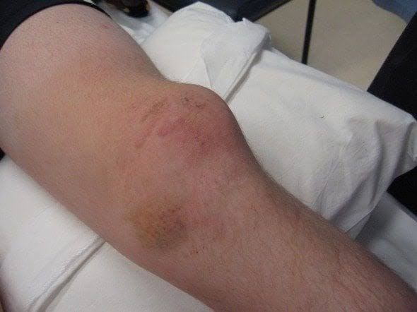
- Medial tenderness: Along the Medial Patellofemoral Ligament (MPFL).
- Patellar Apprehension Test: Positive (pain/fear with lateral force on patella at 20–30° flexion).
Diagnosis
- Initial: Plain Radiographs (AP, Lateral, Sunrise) → r/o osteochondral fractures, confirms reduction.
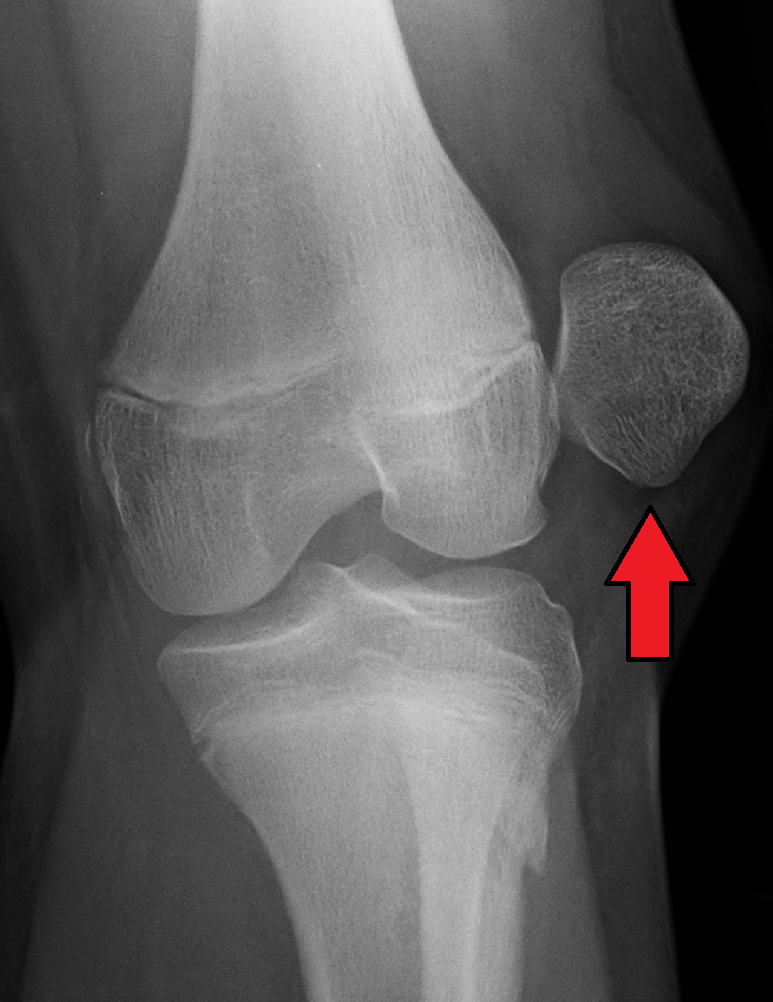
- Confirmatory / Advanced: MRI → gold standard for MPFL tear, chondral lesions, or loose bodies.
- Arthrocentesis: Lipohemarthrosis (fat-fluid levels = intra-articular fracture).
Differential Diagnostics
- ACL Tear: Diff by (+) Lachman/Drawer tests; patella normally positioned; intra-articular pop.
- Tibiofemoral Dislocation: Diff by gross limb deformity, multiligament tears; requires urgent popliteal artery assessment (ABI, CTA).
- Quad / Patellar Tendon Rupture: Diff by inability to straight-leg raise, palpable tendon gap (quad rupture = patella baja; patellar rupture = patella alta).
- Meniscal Tear: Diff by (+) McMurray test, joint line tenderness, delayed joint effusion (24 hrs).
Management
- Acute (First-line):
- Closed Reduction: Medially directed pressure on patella while gently extending the knee.
- Post-reduction X-ray: Rule out associated fractures and verify relocation.
- Conservative Care: Brief immobilization (hinged brace in extension 1–2 wks) → PT focused on VMO strengthening + RICE/NSAIDs.
- Surgical (Second-line / Refractory):
- Indications: Osteochondral fracture / loose bodies, irreducible dislocation, or recurrent instability.
- Procedure: Arthroscopic fragment removal/fixation, MPFL reconstruction.
Complications
- Recurrent dislocation/instability (most common; 30–50%).
- Patellofemoral osteoarthritis.
- Intra-articular loose bodies causing joint locking.