Epidemiology
- Prevalence: second most prevalent skeletal disease after osteoporosis in individuals > 50 years of age
Etiology
Pathophysiology
Overview
Idiopathic disease
- Associated with a high rate of bone remodeling: ↑ RANKL-RANK activity → ↑ NF-κB signaling → ↑ osteoclast activity → ↑ osteoblast activity → formation of disorganized (woven) bone
- The result is that newly remodeled bone is more vascular, and lacks the strength and shape of normal bone. This weak bone can be easily deformed by stress (e.g., weight bearing) and is highly susceptible to fractures. Furthermore, bone remodeling in Paget disease can result in enlargement of bone, which can result in entrapment neuropathies in regions where nerves pass through bony tunnels (e.g., cranial nerve canals, skull base foramina, spinal canal).
Stages of Paget disease
Bone remodeling in Paget disease occurs in three phases, followed by a quiescent stage:
- Lytic phase: ↑ number of osteoclasts appear in bone → ↑ osteoclastic activity → ↑ rate of bone resorption
- Mixed lytic and blastic phase: ↑ osteoclastic activity is accompanied by an ↑ number of osteoblasts, which infiltrate the lacunae → ↑ rate of bone formation with haphazardly laid collagen fibers → formation of abnormal hypervascular woven bone
- Sclerotic phase: Osteoblastic activity overtakes osteoclastic activity, which leads to formation of dense, sclerotic bone.
- Quiescent stage: Both osteoclastic and osteoblastic activity cease (“quiet phase” of the disease).
Disease localization
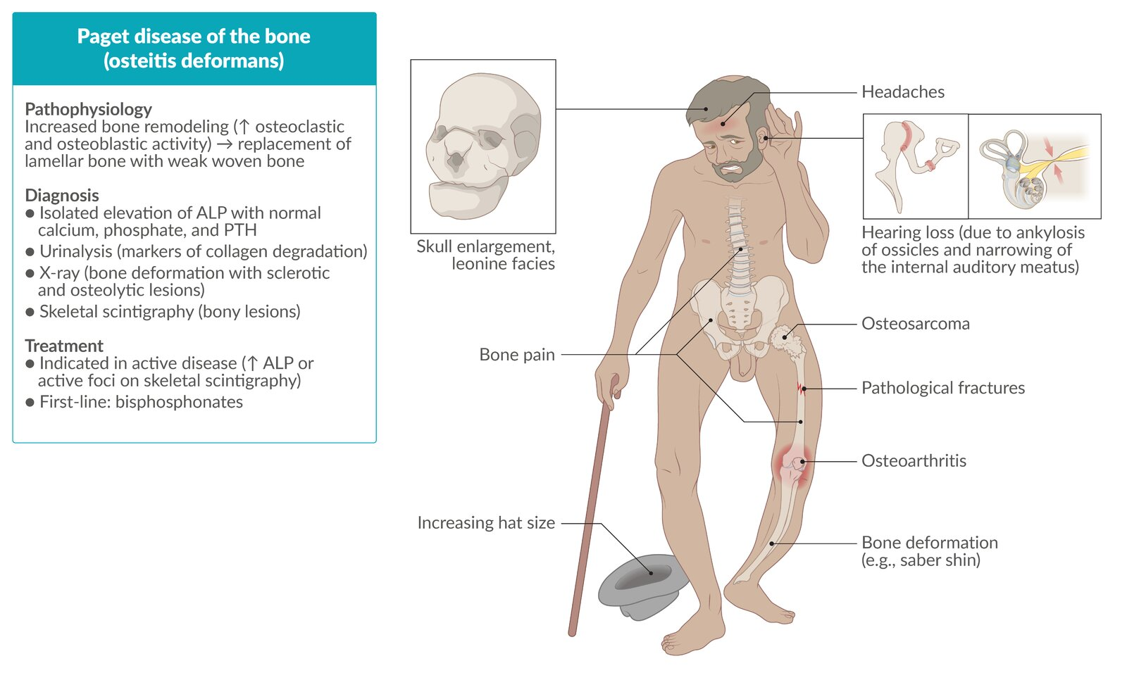
The pelvis, skull, vertebral column, and long bones of the lower extremities are the most commonly affected sites.
Clinical features
- Asymptomatic (70–80%): Incidental finding of isolated markedly elevated ALP or abnormal plain radiograph.
- Skeletal & Joint:
- Bone pain: Dull, deep, aching; persists at rest/night.
- Bone deformities: Bowing of long bones (femur/tibia), kyphosis, increased skeletal volume.
- Local erythema/warmth over affected bones (due to hypervascularity/AV shunting).
- Secondary osteoarthritis of adjacent joints (hip, knee).
- Craniofacial:
- Increasing hat size (frontal bossing, skull enlargement).
- Sensorineural hearing loss (CN VIII compression or cochlear capsule involvement).
- Headaches, dizziness, facial numbness, dental malocclusion.
- Spinal:
- Spinal canal stenosis, radiculopathy, spinal cord compression.
Diagnostics
- Radiographs typically reveal lytic or mixed lytic-sclerotic lesions, thickening of cortical and trabecular bone, and bony deformities.
- Serum total ALP: often significantly increased; first-line test to assess disease severity
- Due to increased osteoblast activity
- Serum calcium, phosphate, and parathyroid hormone (PTH) levels: typically normal c
- Normal homeostasis is usually maintained despite the bone remodeling involved in PDB.
- Elevated bone turnover markers: Serum P1NP, urine CTx/NTx, urine hydroxyproline (breakdown of collagen).
- Bone biopsy
- Typically shows a chaotic, mosaic-like pattern of irregularly juxtaposed lamellar and woven bone
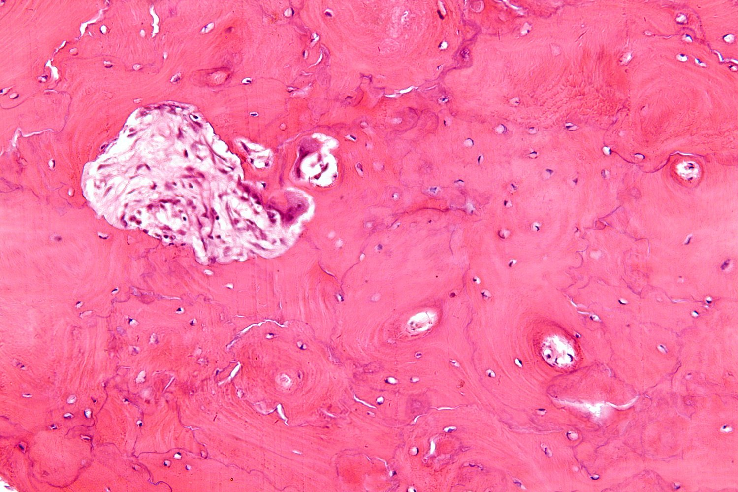
- Typically shows a chaotic, mosaic-like pattern of irregularly juxtaposed lamellar and woven bone
Treatment
Bisphosphonates
- First-line therapy for patients with bone pain and those at risk of bone complications
- Common Agents
- Alendronate, Risedronate (oral weekly)
- Zoledronic acid (IV)
- Mechanism: induce apoptosis of osteoclasts
- Bisphosphonates are pyrophosphate analogs that bind to hydroxyapatite binding sites on the surface of bone tissue: uptake by osteoclasts during phases of bone resorption → interference with osteoclast function and promotion of osteoclast apoptosis → reduced bone resorption
- Indications
- Osteoporosis (1st-line)
- Paget disease
- Hypercalcemia of malignancy
- Bone metastases
- Adverse effects
- Bisphosphonate-related osteonecrosis of the jaw: adverse effect of bisphosphonates, usually seen with high-dose IV administration in tumor patients
- Etiology: Trauma to the jaw (e.g., tooth extractions or other dental procedures) is a trigger in many cases.
- Pathophysiology: not yet fully understood
- Clinical features: chronic swelling, mild pain, exposed necrotic bone
- Treatment: supportive, e.g.,:
- Improving oral hygiene
- Antibiotic rinses
- Limited debridement
- Prevention
- Dental examination in conjunction with treatment of preexisting dental conditions (e.g., extraction of unrestorable teeth) prior to initiation of therapy
- Establishing a daily oral care plan that involves daily oral rinsing, brushing, and flossing
- Esophageal inflammation and cancer
- To avoid this complication, patients should swallow medication with a sufficient amount of water and maintain an upright position for 30 minutes.
- Bisphosphonate-related osteonecrosis of the jaw: adverse effect of bisphosphonates, usually seen with high-dose IV administration in tumor patients
Complications
- Osteosarcoma / Malignant Transformation (< 1%):
- Suspect in pts with sudden worsening of localized pain, swelling, or enlarging mass.
- Imaging shows new destructive lytic lesion, cortical destruction, and Codman triangle / “sunburst” periosteal reaction.
- Very poor prognosis.
- High-Output Heart Failure:
- Caused by extensive arteriovenous (AV) micro-shunting within hypervascularized pagetic bone.
- Signs: Wide pulse pressure, brisk peripheral pulses, S3 gallop, elevated BNP.
- Pathologic Fractures:
- “Chalkstick” fractures (transverse, clean break through brittle, sclerotic long bones).
- Neurological Deficits:
- Sensorineural hearing loss, blindness (optic canal stenosis), paraplegia/cauda equina syndrome.
- Nephrolithiasis / Hypercalciuria:
- Seen primarily during periods of forced immobilization.