Nummular Dermatitis
Epidemiology & Risk Factors
- Older adults (M > F); young adult females.
- Severe xerosis (winter, low humidity, dry climate).
- Frequent hot showers, harsh soaps, atopic diathesis.
Clinical Features
- “Coin-shaped” (discoid), well-demarcated pruritic erythematous plaques (1–5 cm).
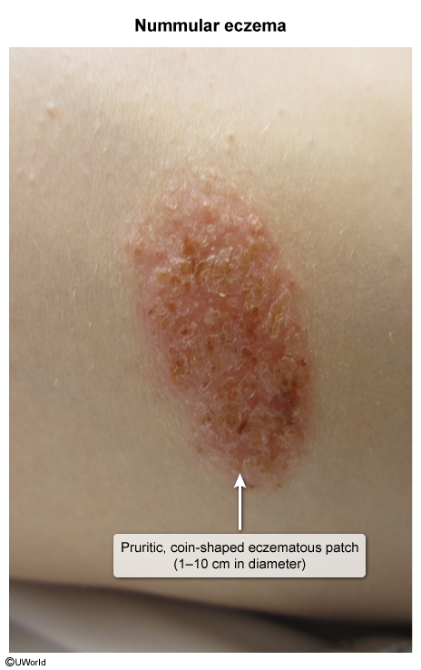
- Location: Extremities (pretibial legs, arms) > trunk. Face/scalp spared.
- Acute: Erythematous papules/vesicles with exudation and crusting.
- Chronic: Dry, scaly, lichenified plaques.
Diagnosis
- Initial/Screening: Clinical diagnosis based on morphology and xerosis.
- Key Labs: KOH prep negative for hyphae (rules out fungal infection).
- Confirmatory/Biopsy: Rarely needed; shows spongiotic dermatitis.
Differential Diagnostics
- Tinea Corporis: Diff by central clearing, advancing scaly border, and (+) KOH prep.
- Plaque Psoriasis: Diff by silvery scale, extensor distribution, and (+) Auspitz sign.
- Contact Dermatitis: Diff by distribution matching specific allergen exposure, (+) patch test.
- Impetigo: Diff by honey-colored crusting, acute onset, (+) bacterial culture.
Management
- First-line:
- High-potency topical corticosteroids (e.g., Clobetasol 0.05%, Triamcinolone 0.1%).
- Aggressive emollients (petrolatum) applied immediately after bathing (“soak and seal”).
- Lifestyle: Lukewarm/brief showers, mild cleansers.
- Second-line: Topical calcineurin inhibitors (Tacrolimus), NB-UVB phototherapy.
- If Superinfected: Oral anti-staphylococcal Abx (Cephalexin).
Complications
- Secondary bacterial impetiginization / Cellulitis (S. aureus).
- Lichen simplex chronicus from chronic scratching.