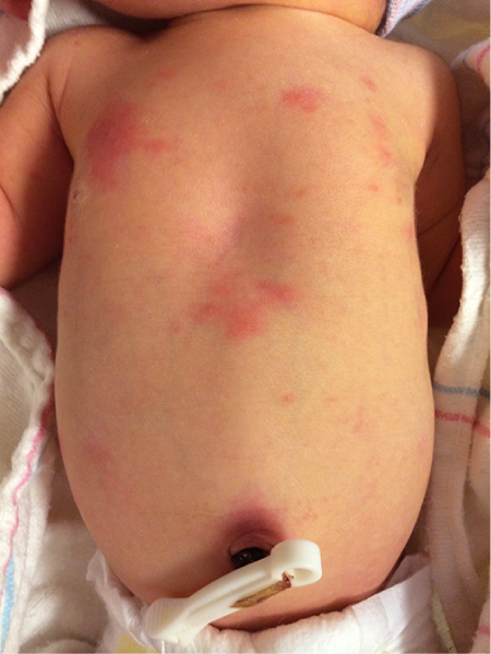
Onset: Day 1–3 of life (peaks at 24–48 hours; rare in premature infants).
Benign, self-limited neonatal rash.
Clinical Features: Blotchy, erythematous macules with central pale papules or pustules (“flea-bitten” appearance); distributed on trunk and proximal extremities; spares palms and soles.
Clinical Features: 1–2 mm pearly white/yellow papules on the nose, cheeks, and forehead. Epstein pearls are identical lesions located on the hard palate/gingiva.
Management: Reassurance; resolves spontaneously within the first few weeks to months.
Miliaria (Heat Rash)
Pathophysiology: Occlusion of eccrine sweat ducts due to overheating/bundling.
Subtypes:
Miliaria Crystallina: Superficial clear vesicles without inflammation (resembles dew drops).
Miliaria Rubra: Pruritic, erythematous papules/pustules in intertriginous/occluded areas.
Onset: Typically at 2–3 weeks of life (distinguishes from ETN/TNPM).
Pathophysiology: Inflammatory reaction to Malassezia colonization; maternal androgens.
Clinical Features: Inflammatory papules and pustules on cheeks and nose; lacks true comedones.
Management: Gentle cleansing w/ soap and water. If severe/refractory: topical 2% ketoconazole or 1% hydrocortisone.
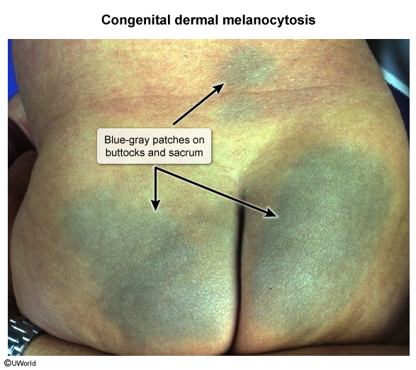
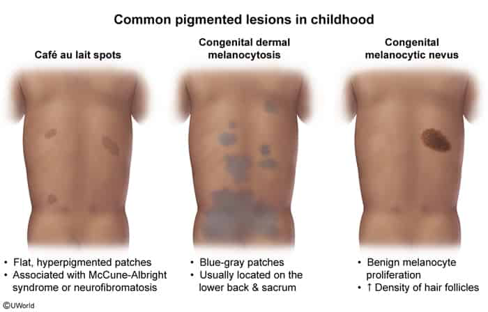
Congenital Dermal Melanocytosis (Mongolian Spot)
Pathophysiology: Failure of embryologic migration of melanocytes from the neural crest to the epidermis; arrested melanocytes reside deep within the dermis (Tyndall effect causes blue-gray appearance).
Epidemiology: Extremely common in neonates of Asian, African, Hispanic, and Native American descent (80–90%).
Clinical Features:
Flat, poorly demarcated, blue-gray or slate-brown macules and patches.
Location: Most commonly the lumbosacral region and buttocks; occasionally on shoulders, back, or posterior thighs.
Normal skin texture: No induration, tenderness, or edema.
Diagnosis: Purely clinical.
Differential Diagnosis:
Non-accidental trauma / Child abuse (Bruising): Bruises are tender, have overlying swelling, change color over days (red/purple → yellow/green), and occur in atypical locations. Dermal melanocytosis remains stable, non-tender, and uniform in color.
Nevus of Ota: Dermal melanocytosis along the ophthalmic (V1) and maxillary (V2) divisions of CN V; involves sclera and periorbital skin.
Nevus of Ito: Dermal melanocytosis in the supraclavicular/deltoid and scapular distribution.
Management:
Reassurance: Benign condition with no malignant potential. c
Documentation: Must be meticulously documented in the neonatal medical record at birth to prevent future false suspicion or allegations of physical child abuse.
Course: Gradually fades over the first few years of life; usually resolves completely by puberty.
Congenital Melanocytic Nevus (CMN)
Pathophysiology: Benign proliferation of neural crest-derived melanocytes present at birth.
Clinical Features:
Hyperpigmented, well-circumscribed macule, patch, or plaque.
Surface may become verrucous, rugose, or pebbly with age.
Size Stratification (Projected Adult Size - PAS):
Small: < 1.5 cm.
Medium: 1.5 – 19.9 cm.
Large / Giant: ≥ 20 cm (or > 5% total body surface area; classic “bathing trunk” or “garment” distribution).
Complications & High-Yield Associations:
Malignant Melanoma: Risk correlates directly with size. Giant CMN carries a 2–5% lifetime risk of malignant melanoma (highest in the first decade of life). Small/medium lesions carry minimal/negligible risk.
Neurocutaneous Melanocytosis (NCM): Melanocyte deposition in the leptomeninges and brain parenchyma.
Risk Factors: Giant CMN (especially on axial/posterior trunk or head/neck) or ≥ 3 satellite nevi.
Screening: Brain and Spine MRI w/ contrast (ideally performed before 4–6 months of age, prior to complete normal brain myelination which obscures leptomeningeal lesions) for infants with giant CMN or high-risk features (multiple satellite nevi, axial location).
Regular dermatologic surveillance w/ serial photography and dermoscopy.
Management:
Small/Medium: Serial observation and routine dermatologic monitoring.
Giant/Large: Surgical excision (staged excision or tissue expansion) considered for cosmesis or atypical changes. Note: Surgical removal reduces but does not entirely eliminate the risk of melanoma, as malignant transformation can occur in deeper subcutaneous tissue or the central nervous system.