Caused by Molluscum Contagiosum Virus (MCV), a member of the poxvirus family.
Transmission: direct skin-to-skin contact, autoinoculation (scratching), fomites, or sexual contact (adults).
Risk Factors:
Children (especially school-aged, due to close contact and swimming pools).
Atopic dermatitis (AD) due to impaired skin barrier facilitating spread.
Immunocompromised state (especially HIV with CD4 < 200). c
Pathophysiology
Clinical features
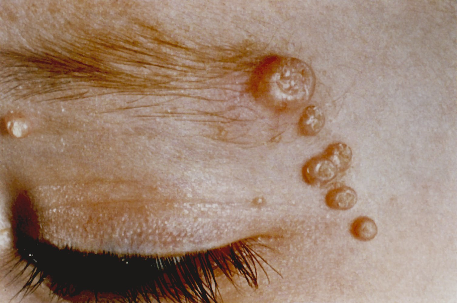
Morphology: Flesh-colored, pearly, dome-shaped papules with characteristic central umbilication. c2
Predilection sites:
In children: face, trunk, and extremities (e.g., axilla, antecubital and popliteal fossa)
In adults: lower abdomen, groin, genitalia, and proximal thighs
Diagnostics
Dx is typically made clinically based on the pathognomonic appearance of the lesions.
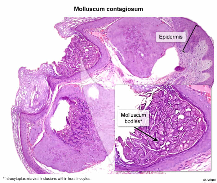
Histology is diagnostic and shows characteristic large, intracytoplasmic eosinophilic inclusion bodies, known as Henderson-Paterson bodies or molluscum bodies, within keratinocytes.
The epidermis shows cup-shaped, inverted lobular hyperplasia.
Treatment
First-line (Healthy Children): Reassurance & observation (spontaneous resolution typically occurs within 6–12 months). c
Active Intervention (Indicated for severe pruritus, rapidly spreading lesions, cosmetic concern, or sexual transmission in adults):