Epidemiology & Risk Factors
Demographics :
Young, active athletes: Acute traumatic injuries (rotational force on a planted foot).
Older adults (>40-50 yo): Degenerative tears with minimal/trivial trauma (e.g., squatting, rising from a chair).
Anatomy :
Medial meniscus > lateral meniscus tears (medial is less mobile and anchored to the MCL).Associated with concurrent ACL tears (e.g., “unhappy triad”: ACL, MCL, medial meniscus).
Clinical Features
History :
Acute onset with a twisting mechanism on a flexed, weight-bearing knee.
Sensation of a “pop” followed by delayed joint swelling/effusion (develops over 12–24 hours; vs immediate hemarthrosis in ACL tears).
Mechanical symptoms: Intermittent catching, clicking, or locking ; feeling of knee “giving way” or instability.
Physical Exam :
Joint-line tenderness : High sensitivity; localized to the medial or lateral joint line.Effusion/mild joint swelling.
Provocative Maneuvers :
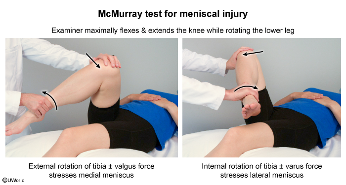
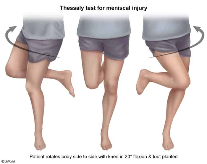
McMurray test : Palpable or audible “click” / pain during passive knee flexion-extension with internal/external rotation.Thessaly test : Pain or catching while standing on the affected leg (flexed at 20°) and rotating the knee internally and externally.Apley grind test : Pain on axial compression and tibial rotation with pt in prone position (flexed 90°).
Diagnosis
Initial / Screening :
Plain Radiographs (XR) (AP, lateral, sunrise, and weight-bearing views):
Usually normal in acute tears.
Performed first to rule out fractures, loose bodies, or underlying OA.
Confirmatory / Non-invasive Gold Standard :
MRI of the knee : Test of choice; demonstrates high-signal intensity extending through the meniscal surface.
Definitive (Invasive) :
Diagnostic Arthroscopy : Reserved for cases planned for simultaneous therapeutic intervention.
Differential Diagnostics
ACL Tear :
Diff by: Immediate gross hemarthrosis (within minutes to hours), rapid onset swelling, (+) Lachman test (most sensitive), (+) Anterior drawer test.
MCL Tear :
Diff by: Mechanism of pure valgus stress, localized pain at the medial femoral epicondyle, laxity with valgus stress testing at 30° flexion, no mechanical locking.
Patellofemoral Pain Syndrome (PFPS) :
Diff by: Chronic anterior retropatellar pain aggravated by prolonged sitting (“theater sign”) or stair descent, no joint-line tenderness, no true effusion.
Pes Anserine Bursitis :
Diff by: Pain localized inferior to the medial joint line over the anteromedial proximal tibia, normal range of motion (ROM), no mechanical locking or intra-articular effusion.
Knee Osteoarthritis (OA) :
Diff by: Insidious onset, age >50 yo, morning stiffness <30 min, crepitus, XR showing asymmetric joint space narrowing and osteophytes.
Management
First-line (Conservative Therapy) :
Indications : Degenerative tears in older pts, mild/infrequent symptoms, stable tears without mechanical locking.Regimen :
RICE (Rest, Ice, Compression, Elevation) + activity modification.NSAIDs for pain/inflammation.Physical therapy (PT) focused on quadriceps and hamstring strengthening.
Second-line / Surgical Intervention :
Indications : Persistent symptoms after 4–6 weeks of conservative therapy, younger/active athletic pts with acute tears, or persistent “locked knee” (mechanical obstruction).Procedures :
Arthroscopic Meniscal Repair : Preferred for peripheral (“red-red” zone) tears due to good vascularity and high healing potential.Partial Meniscectomy : Performed for avascular (“white-white” central zone) or complex/macerated tears that cannot heal.
Complications
Early accelerated knee osteoarthritis (esp. post-meniscectomy due to altered load transmission).
Chronic articular cartilage erosion/chondral defects.
Baker cyst (popliteal cyst) formation secondary to chronic joint effusion.Arthrofibrosis or persistent joint stiffness post-op.