Enthesitis-related (HLA-B27 associated) and Psoriatic JIA.
Clinical Features
General: Joint pain, swelling, warmth, morning stiffness that improves with activity, and limp without refusal to bear weight. c2
Oligoarticular JIA:
Asymmetric involvement of large joints (knee > ankle > wrist > elbow).
Typically painless or mildly painful; child may present with a limp.
High risk of asymptomatic anterior uveitis (insidious, leads to blindness if missed).
Polyarticular JIA:
Symmetric involvement of small and large joints (hands, feet, knees, cervical spine).
Resembles adult RA; may have low-grade fever and mild anemia.
Systemic JIA (sJIA):
Quotidian fever: Daily, high-spiking fever (> 38.5°C) typically occurring once/twice daily in the late afternoon/evening, returning to baseline. c
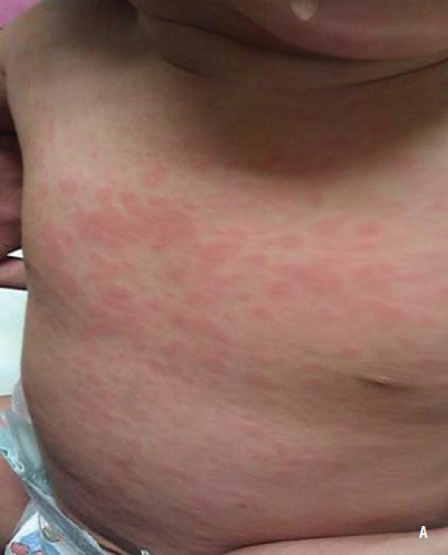
Evanescent salmon-pink macular rash: Coincides with fever spikes; non-pruritic, transient, mostly on trunk/extremities. c
Hepatosplenomegaly, generalized lymphadenopathy, and serositis (pericardial/pleural effusions).
Polyarthritis (often develops weeks to months after systemic features).
Diagnosis
Clinical diagnosis of exclusion (age < 16 yo, persistent joint swelling/effusion ≥ 6 weeks, other causes ruled out).
Key Labs:
Inflammatory markers: ↑ ESR, ↑ CRP, thrombocytosis, leukocytosis, microcytic anemia of chronic disease. c
sJIA Labs: Extreme leukocytosis (WBC > 20,000-30,000/mm³), marked thrombocytosis, and dramatically elevated serum ferritin. c
Autoantibodies:
ANA: Positive in ~60-80% of oligoarticular JIA (strongly associated with risk of anterior uveitis; does not correlate with disease activity).
RF / Anti-CCP: Checked in polyarticular JIA (determines prognosis; RF (+) carries worse prognosis). Negative in sJIA.
Screening:
Regular Slit-Lamp Examination: Mandatory at diagnosis and scheduled every 3–6 months for all oligoarticular/polyarticular pts (especially ANA(+)) to screen for silent anterior uveitis.
Imaging:
Plain X-ray (Initial): Soft tissue swelling, osteopenia, accelerated epiphyseal growth (early); joint space narrowing and erosions (late).
Ultrasound / MRI: Detects subclinical synovitis, tenosynovitis, and cartilage loss.
Arthrocentesis (if monoarticular): Performed to rule out septic arthritis (JIA synovial fluid: WBC 5,000–50,000/mm³ with moderate PMN predominance; cultures negative).
Differential Diagnostics
Septic Arthritis:
Diff by: Acute onset, single joint, high fever, severe pain with refusal to bear weight, synovial WBC > 50,000/mm³ with > 75% PMNs, (+) Gram stain/culture.
Transient Synovitis:
Diff by: Acute hip pain following viral URI in a child aged 3–8 yo, self-limiting within 1–2 weeks, normal to mildly elevated ESR/CRP.
Acute Lymphoblastic Leukemia (ALL):
Diff by: Severe bone/joint pain (worse at night, waking from sleep), out of proportion to physical exam findings, cytopenias (anemia, thrombocytopenia, leukopenia), peripheral blasts.
Acute Rheumatic Fever (ARF):
Diff by: Migratory polyarthritis (moves from joint to joint), preceding group A Streptococcus pharyngitis, carditis, Sydenham chorea, erythema marginatum, elevated ASO/anti-DNase B titers.
Lyme Arthritis:
Diff by: History of tick bite/travel to endemic areas, prominent knee effusion with minimal pain, positive Borrelia burgdorferi serology (ELISA followed by Western blot).