Achilles tendinopathy

  • Epidemiology & Risk Factors
    • Overuse/running/jumping sports; sudden increase in training volume/intensity.
    • Medications: Fluoroquinolones (e.g., ciprofloxacin), systemic corticosteroids.
    • Associated conditions: Seronegative spondyloarthropathies (enthesitis), familial hypercholesterolemia (xanthomas), DM.
  • Pathophysiology
    • Degenerative tendinosis: Repetitive mechanical overload exceeding repair capacity leads to failed healing (predominantly degenerative, non-inflammatory tissue response).
    • Collagen disruption: Normal parallel Type I collagen is degraded and replaced by weaker, disorganized Type III collagen with increased proteoglycans/mucoid matrix.
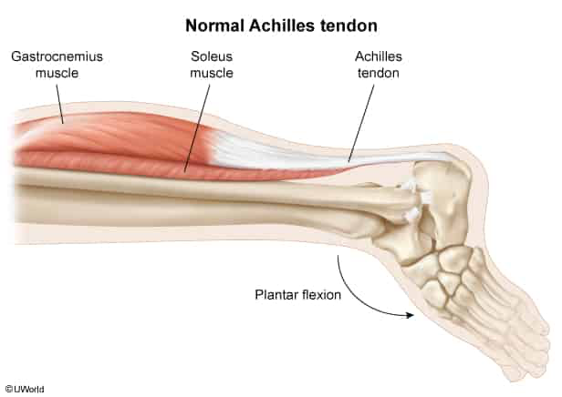
    • Vascular watershed area: Relative hypovascularity 2–6 cm proximal to calcaneal insertion predisposes to micro-ischemia, tissue breakdown, and impaired healing.
  • Clinical Features
    • Burning posterior heel pain and stiffness (worse in AM and after exercise; improves with light activity).
    • Midportion tendinopathy (most common): Tenderness/swelling 2–6 cm proximal to calcaneal insertion.
    • Insertional tendinopathy: Pain directly at calcaneal attachment ± Haglund deformity.
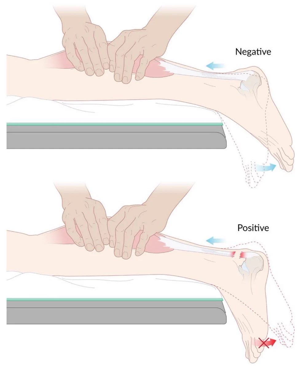
    • Negative Thompson test (intact plantarflexion on calf squeeze).
  • Diagnosis
    • Initial / Primary: Clinical diagnosis via H&P.
    • Ultrasound (US): Thickened tendon, hypoechoic degenerative areas, neovascularization.
    • MRI (Gold Standard / Confirmatory): Increased T1/T2 signal; evaluates partial vs. full-thickness tears.
    • X-ray: Normal or shows calcaneal spur/calcification (rules out avulsion or stress fracture).
  • Differential Diagnostics
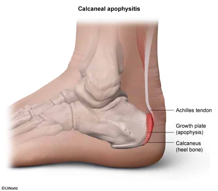
    • Calcaneal Apophysitis (Sever Disease): Diff by occurrence in skeletally immature pediatric/adolescent athletes (ages 8–12) during growth spurts; pain localized over the posterior calcaneal apophysis with (+) calcaneal squeeze test.
    • Achilles Tendon Rupture: Sudden “pop” or “gunshot” sensation, palpable gap, (+) Thompson test.
    • Plantar Fasciitis: Inferior/plantar heel pain, worse with first morning steps.
    • Calcaneal Stress Fracture: Pain with calcaneal squeeze test (medial-lateral compression).
    • Retrocalcaneal Bursitis: Pain on two-finger palpation anterior to the Achilles insertion.
  • Management
    • First-line:
      • Relative rest, activity modification, ice, temporary heel lifts.
      • Physical therapy: Eccentric loading exercises (mainstay of rehab). c
      • Short-term oral NSAIDs for acute pain control.
    • High-Yield Contraindication: NO local glucocorticoid injections (high risk of tendon rupture).
    • Refractory (>3–6 months): Extracorporeal shock wave therapy (ESWT) or surgical debridement.
  • Complications
    • Achilles tendon rupture (partial or complete).
    • Chronic debilitating pain and altered gait biomechanics.

Iliotibial band syndrome

  • Etiology & Risk Factors:
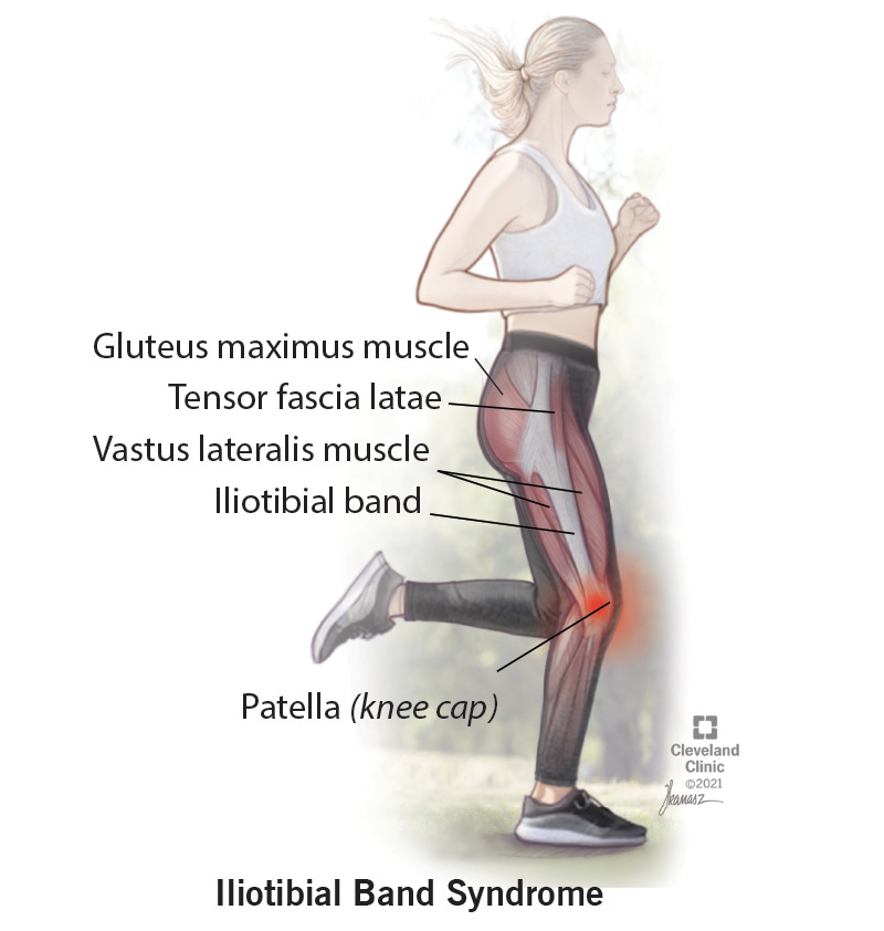
    • Repetitive friction of the distal ITB against the lateral femoral epicondyle (~30° knee flexion).
    • Common in: Distance runners, cyclists, military recruits.
    • Biomechanical trigger: Weak hip abductors (gluteus medius) causing pelvic drop and increased ITB tension; running on banked/downhill surfaces.
  • Clinical Features:
    • Sharp, burning lateral knee pain worsening with repetitive foot-strikes/downhill running.
    • Pain relieved by rest; no joint effusion and no mechanical symptoms (no true locking/catching).
  • Physical Exam Findings:
    • Point tenderness over the lateral femoral epicondyle (~2–3 cm proximal to lateral joint line).
    • (+) Noble test: Pain reproduced at ~30° flexion while compressing the lateral femoral epicondyle.
    • (+) Ober test: Inability to adduct hip past midline with knee flexed in lateral decubitus (confirms ITB tightness).
  • Diagnosis:
    • Clinical diagnosis based on typical Hx and localized epicondylar tenderness.
    • Imaging (X-ray/MRI) not routinely required; reserved only to rule out intra-articular pathology if atypical.
  • Key Differentials:
    • Lateral meniscal tear: Tenderness at lateral joint line (not epicondyle), (+) joint effusion, (+) McMurray test, mechanical catching/locking.
    • LCL sprain: Acute varus trauma, laxity on varus stress testing.
    • Patellofemoral pain syndrome: Anterior/peripatellar pain, worse with prolonged sitting (“theater sign”).
  • Management:
    • 1st-line (Conservative):
      • Activity modification (relative rest, avoid downhill running), cryotherapy, short course of NSAIDs.
      • Physical Therapy: Strengthening of hip abductors (gluteus medius) + ITB/tensor fasciae latae stretching.
    • 2nd-line (Persistent pain > 4–6 weeks):
      • Local corticosteroid injection deep to the ITB at the lateral femoral epicondyle.
    • Refractory (Rare, > 6 months failed therapy):
      • Surgical ITB release or lateral bursectomy.

Lateral epicondylitis (Tennis elbow)

  • Etiology & Pathophysiology:
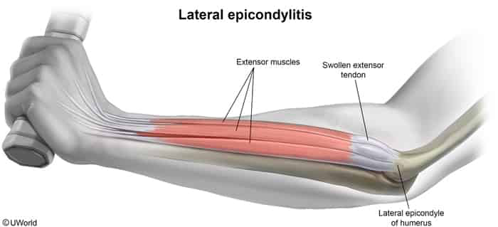
    • Overuse tendinopathy (angiofibroblastic degeneration) of the extensor carpi radialis brevis (ECRB) origin.
    • Triggered by repetitive, forceful wrist extension and supination (e.g., tennis backhand, screwdriver use).
  • Clinical Presentation:
    • Point tenderness localized to the lateral epicondyle.
    • Pain reproduced with resisted wrist extension (with elbow extended).
    • Pain reproduced with resisted middle-finger extension.
    • Pain reproduced with passive wrist flexion.
    • Normal elbow ROM and intact neurovascular exam.
  • Diagnosis:
    • Clinical diagnosis (no labs or imaging needed routinely).
    • XR: Indicated only to rule out bony pathology, OA, or calcific tendinitis.
    • MRI / US: Reserved for atypical presentations or refractory cases prior to surgery (shows ECRB thickening/tears).
  • Key Differentials:
    • Radial tunnel syndrome: Maximal tenderness is 4–5 cm distal to the lateral epicondyle over the mobile wad (PIN compression); lacks epicondyle point tenderness.
    • Cervical radiculopathy (C6–C7): Radiating neck pain, dermatomal paresthesias, diminished reflexes, and (+) Spurling sign.
    • Elbow osteoarthritis: Decreased active/passive elbow ROM, joint line tenderness, and osteophytes on XR.
  • Management:
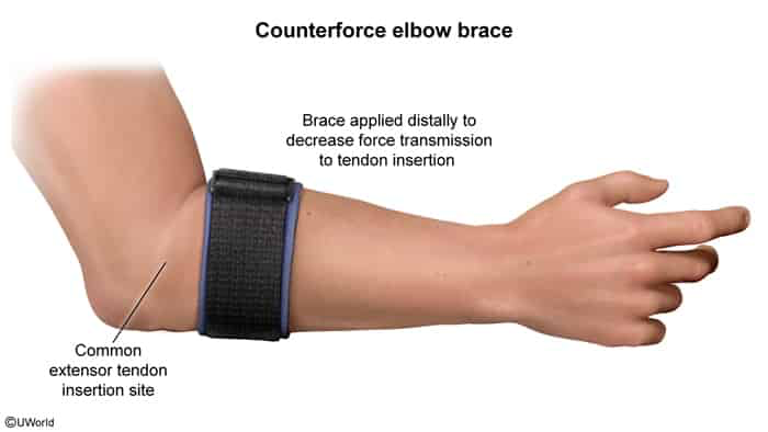
    • First-line (Conservative): Activity modification, short-term NSAIDs, eccentric strengthening PT, and a counterforce elbow strap (forearm band).
    • Second-line: Local corticosteroid injection (provides rapid short-term relief, but high recurrence rate and risk of tendon rupture).
    • Refractory (failed 6–12 months of conservative care): Surgical debridement and release of the ECRB origin.
  • Complications:
    • Chronic lateral elbow pain and reduced grip strength.
    • Iatrogenic tendon rupture or subcutaneous fat atrophy/depigmentation from corticosteroid injections.