Epidemiology


  • Incidence: peak at 10–20 years
    • Second most common primary bone tumor in children and adolescents, after osteosarcoma

Etiology

  • Cell of Origin: Neural crest / neuroectodermal origin (part of the Ewing family of tumors / PNET spectrum).
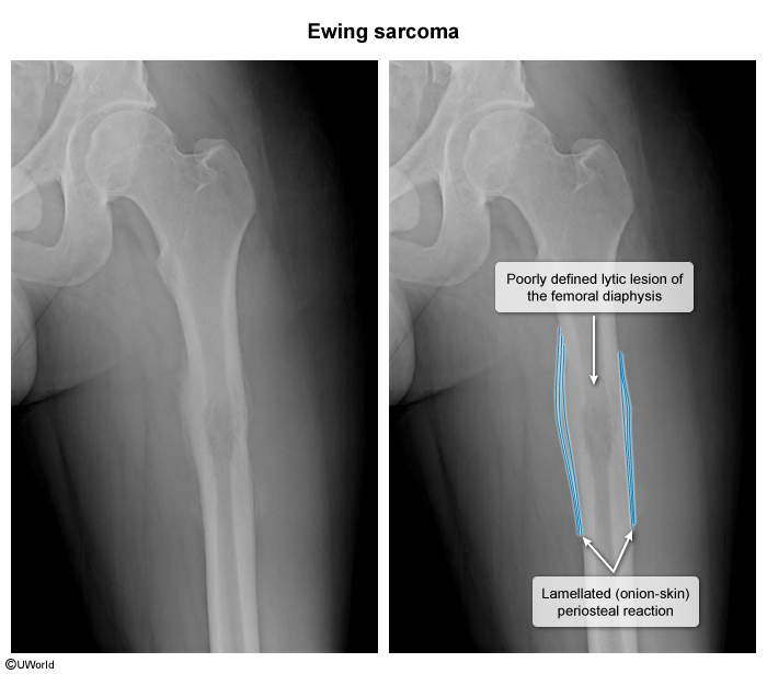
    • Therefore, it is not associated with new bone formation (ie, radiodense areas on X-ray) and is typically located in the diaphysis (vs the metaphysis) c

Pathophysiology


Clinical features


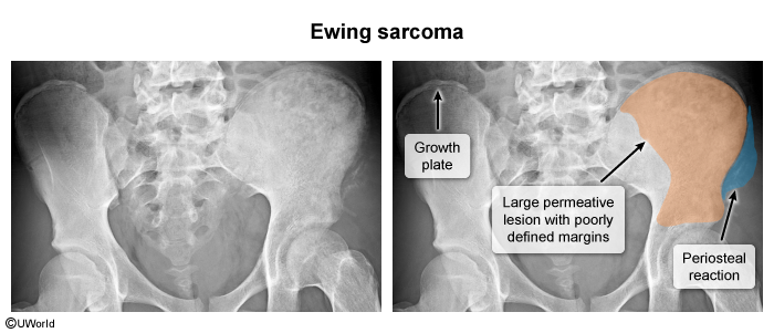
  • Location: Diaphysis and metadiaphysis of long bones (femur > tibia > humerus) and flat/axial bones (pelvis, ribs, spine).
  • Local Symptoms:
    • Localized, deep, progressive bone pain (worse at night or with weight-bearing).
    • Tender, palpable, warm soft-tissue mass overlying the bone.
  • Systemic Symptoms:
    • Fever, malaise, weight loss, night sweats (mimics osteomyelitis or systemic infection). c

Diagnostics


  • Initial Step: Plain Radiograph (X-ray) of the affected extremity.
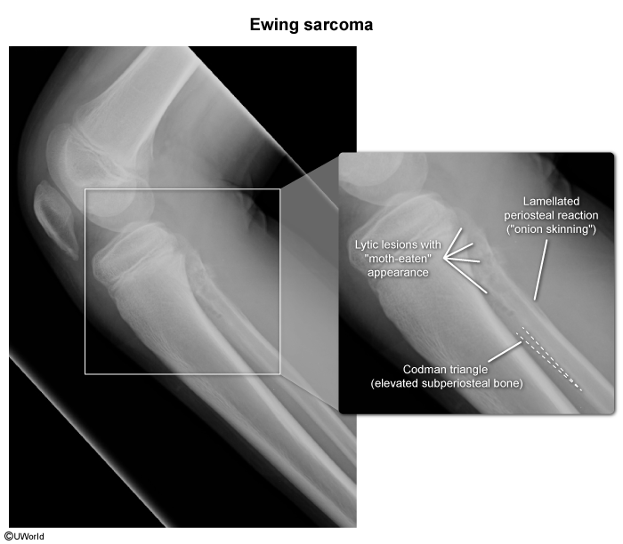
    • Findings: Permeative, osteolytic lesion with poorly defined (“moth-eaten”) margins; soft-tissue mass. c
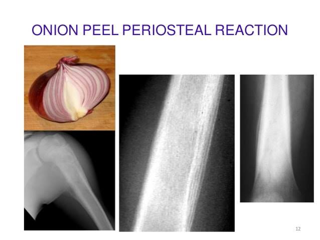
    • Periosteal reaction: Characteristic “onion-skin” (lamellated, multilayered subperiosteal bone formation); may also show Codman triangle.
  • Biopsy
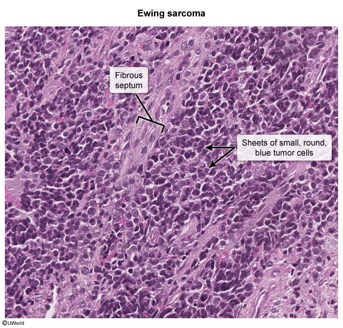
    • Sheets of uniform, small, round, blue cells. c
    • Scant cytoplasm, prominent nuclei.
    • Homer-Wright rosettes (circular grouping of tumor cells around a central fibrillary space) may be present, indicating neural differentiation.
  • Chromosomal translocation t(11;22)(q24;q12) which leads to expression of fusion protein EWS-FLI1

Differential Diagnostics

  • Osteomyelitis:
    • Diff by: Positive blood/bone aspirate cultures, focal cortical destruction without large soft tissue mass, rapid clinical response to targeted Abx.
  • Osteosarcoma:
    • Diff by: Metaphysis location (esp. around the knee: distal femur / proximal tibia), “sunburst” periosteal pattern, pleomorphic spindle cells forming osteoid matrix, rare systemic B-symptoms.
  • Osteoid Osteoma:
    • Diff by: Pain strictly worse at night and rapidly relieved by NSAIDs; XR shows small radiolucent nidus (<1.5–2 cm) surrounded by dense sclerotic bone; no soft tissue mass.
  • Langerhans Cell Histiocytosis (Eosinophilic Granuloma):
    • Diff by: Well-demarcated “punched-out” lytic bone lesions without significant periosteal reaction; biopsy shows CD1a/S100 (+) and Birbeck granules (tennis racket appearance on EM).
  • Neuroblastoma:
    • Diff by: Typically presents in younger children (<2–5 yo); primary adrenal/retroperitoneal mass; ↑ urine catecholamines (VMA, HVA).

Treatment