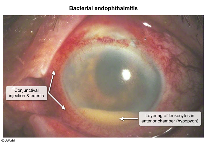
Epidemiology & Risk Factors
Exogenous (most common):
Postoperative ocular surgery : Cataract extraction (most common trigger, typically within 1–2 weeks post-op).Trauma: Penetrating ocular injury, retained intraocular foreign body.
Intravitreal injections (e.g., anti-VEGF for AMD).
Endogenous :
Hematogenous spread from distant focus (e.g., infective endocarditis, indwelling CVC, IVDU).
Immunocompromised status, prolonged TPN.
Common Pathogens :
Coagulase-negative Staphylococci (e.g., S. epidermidis — most common post-op).Staphylococcus aureus , Streptococcus spp.Gram-negative bacilli (e.g., Pseudomonas aeruginosa ).
Candida albicans (most common endogenous agent, especially in IVDU/TPN).
Clinical Features
Symptoms :
Rapidly progressive visual loss / decreased visual acuity (VA).
Significant ocular pain and photophobia (can be mild in indolent coag-negative Staph).
Redness, floaters, eyelid swelling.
Physical Exam :
Hypopyon (layering of WBCs in anterior chamber).Vitritis : Vitreous haziness, loss of red reflex , obscured fundus visualization.Conjunctival injection and chemosis.
Corneal edema, ciliary flush.
Endogenous Candida : Funduscopic exam reveals focal white, fluffy, chorioretinal lesions extending into the vitreous (“string-of-pearls” appearance).
Diagnosis
Initial / Clinical :
Urgent slit-lamp biomicroscopy & dilated fundus exam demonstrating anterior chamber reaction and vitreous exudates.
Confirmatory / Gold Standard :
Vitreous tap / biopsy (or anterior chamber paracentesis): Gram stain, bacterial/fungal cultures, and PCR.
Key Labs :
Blood cultures (mandatory if endogenous source suspected).
Fungal biomarkers (e.g., 1,3-β-D-glucan) if endogenous fungemia suspected.
Imaging :
B-scan ocular ultrasound : Indicated when dense vitritis/corneal edema precludes direct fundus visualization (evaluates vitreous debris, choroidal thickening, retinal detachment).
Differential Diagnostics
Toxic Anterior Segment Syndrome (TASS) :
Diff: Sterile post-op inflammatory reaction presenting within 12–24 hours (vs. days to weeks in endophthalmitis); vitreous is clear; responds rapidly to topical steroids.
Acute Anterior Uveitis (Iritis) :
Diff: Leukocytes confined to anterior chamber; normal red reflex and clear vitreous; no history of intraocular surgery/trauma.
Acute Angle-Closure Glaucoma :
Diff: Markedly elevated IOP (>40–50 mmHg), fixed mid-dilated pupil, “steamy” cornea, headache/nausea; absent hypopyon/vitritis.
Bacterial Keratitis (Corneal Ulcer) :
Diff: Infection limited to corneal stroma with overlying epithelial defect visible on fluorescein staining ; intraocular vitreous initially uninvolved.
Orbital Cellulitis :
Diff: Extraocular muscle restriction, painful ophthalmoplegia, proptosis; internal eye structures (vitreous/retina) are clear.
Management
Immediate Step :
Emergent Ophthalmology consultation (vision-threatening emergency).
First-line Medical Therapy :
Intravitreal empiric broad-spectrum antibiotics :
Vancomycin (Gram-positive coverage) + Ceftazidime or Amikacin (Gram-negative coverage).
If fungal etiology suspected/confirmed:
Intravitreal Amphotericin B or Voriconazole + systemic antifungals (e.g., IV/PO Voriconazole or Fluconazole).
Adjunctive therapy: Topical cycloplegics (for ciliary spasm/pain) and delayed topical corticosteroids (to reduce inflammatory damage after Abx initiated).
Procedural / Refractory :
Pars Plana Vitrectomy (PPV) :
Indicated immediately for patients with profound vision loss (light perception only or worse, per EVS guidelines).
Indicated for retained intraocular foreign body, fungal endophthalmitis, or clinical deterioration despite intravitreal Abx.
Complications
Permanent vision loss / blindness.
Retinal detachment.
Panophthalmitis (extension beyond intraocular space to entire eyeball/orbit).Phthisis bulbi (atrophic, non-functioning, shrunken eye).
Enucleation or evisceration.