Auricular hematoma
- Etiology & Pathophysiology:
- Blunt shearing trauma to the external ear (classic in wrestling, boxing, rugby).
- Separation of perichondrium from underlying cartilage → subperichondrial hematoma → cartilage ischemia and avascular necrosis.
- Clinical Presentation:
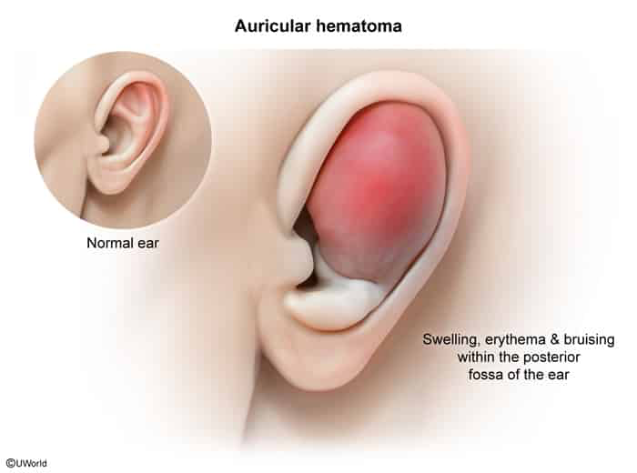
- Acute, tender, tense, fluctuant swelling over the anterior pinna (typically scaphoid fossa or concha).
- Loss of normal cartilaginous architecture and ear landmarks.
- Diagnosis:
- Clinical diagnosis (no labs/imaging routinely required).
- Non-contrast CT indicated only if concomitant skull base or temporal bone fracture is suspected.
- Management:
- Prompt drainage (< 24–48 h): Incision & Drainage (I&D) is the preferred treatment (needle aspiration carries high recurrence; reserved for < 2 cm). c
- Bolster / Pressure dressing: Mandatory post-drainage step for 3–7 days to re-approximate perichondrium to cartilage and prevent reaccumulation.
- Empiric PO Abx: Coverage for skin flora and Pseudomonas aeruginosa (e.g., ciprofloxacin or amoxicillin-clavulanate).
- No return to contact sports until completely resolved (minimum 1–2 weeks).
- Key Complications:
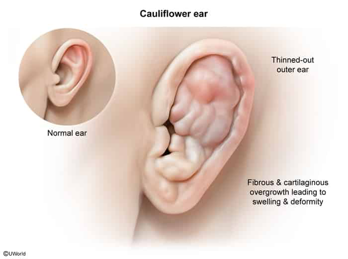
- “Cauliflower ear” (permanent deformity due to neocartilage formation and avascular necrosis).
- Suppurative chondritis / abscess (often Pseudomonas or S. aureus).
- Recurrent hematoma (highest risk if bolster dressing is omitted).