- Spondylosis: a broad term used to describe degenerative changes of the spine that may result in irritation and/or damage of the adjacent nerve roots or spinal cord
Epidemiology
Etiology
- Risk Factors
- Age (Most significant factor).
- Smoking (Vasoconstriction leads to hypoxia and malnutrition of the avascular disc). t
- Obesity (Increased mechanical load).
- Genetics (Family history/predisposition).
- Occupation (Heavy lifting, repetitive bending, vibration).
- Trauma (Previous spinal injury).
Pathophysiology
Clinical features
- Axial Pain: Most common symptom. Localized, chronic, aching pain in the neck (cervical) or low back (lumbar).
- Typically worse with prolonged sitting, bending, or lifting.
- Often relieved by changing positions or lying down.
- Radiculopathy: Nerve root compression causing pain, paresthesia, or weakness in a specific dermatomal/myotomal distribution.
- Lumbar: Sciatica is common (L4-S1 radiculopathy). Provoked by a positive straight leg raise test.
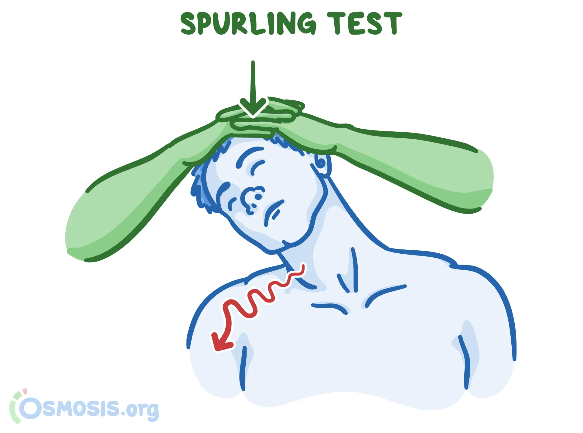
- Cervical: Pain/numbness radiating into the shoulder, arm, or hand. Provoked by a positive Spurling’s test (reproducing symptoms by extending and rotating the neck toward the affected side).
- Myelopathy: Spinal cord compression (most common in the cervical spine). Presents with upper motor neuron (UMN) signs below the level of compression:
- Spasticity, hyperreflexia, positive Babinski sign.
- Gait instability, poor fine motor control in hands, and potential bowel/bladder dysfunction.
Cervical radiculopathy
- Etiology:
- Herniated nucleus pulposus (HNP): Most common in pts <50 yo.
- Cervical spondylosis (osteophytes/foraminal stenosis): Most common in pts >50 yo.
- Most common nerve roots: C7 (most common, ~60–70%) > C6 (~20%).
- High-Yield Root Localization:
- C5: Deltoid/biceps weakness; sensory loss over lateral shoulder/upper arm; ↓ Biceps reflex.
- C6: Biceps/brachioradialis & wrist extension weakness; sensory loss over thumb & index finger; ↓ Brachioradialis & Biceps reflexes.
- C7 (Most common): Triceps & wrist/finger extension weakness; sensory loss over middle finger; ↓ Triceps reflex.
- C8: Finger flexion/intrinsic hand weakness; sensory loss over 4th & 5th digits; no reflex change.
- Key Physical Exam Signs:
- (+) Spurling test: Neck extension + ipsilateral rotation + axial load reproduces radicular arm pain (high specificity).
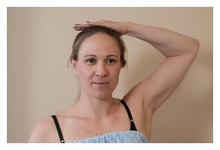
- (+) Shoulder abduction test (Bakody sign): Arm placed over head relieves pain (decreases root tension).
- Lower Motor Neuron (LMN) pattern: Flaccid weakness, hyporeflexia, dermatomal sensory loss; no UMN signs (rules out myelopathy).
- (+) Spurling test: Neck extension + ipsilateral rotation + axial load reproduces radicular arm pain (high specificity).
- Diagnostic Strategy:
- Initial: Clinical diagnosis if mild/moderate symptoms and no “red flags.”
- Confirmatory / Best test: MRI cervical spine.
- Indications: Progressive/severe motor deficit, suspected malignancy/infection/fracture, or symptoms refractory to 4–6 weeks of conservative care.
- EMG/NCS: Reserved for equivocal cases to rule out peripheral entrapment (e.g., CTS, cubital tunnel).
- Management:
- 1st-line (Conservative): NSAIDs / acetaminophen + PT + activity modification ± short-term oral steroid burst.
- 2nd-line (Interventional): Cervical epidural steroid injection (ESI) for persistent pain refractory to 4–6 weeks of conservative therapy.
- Surgical decompression (ACDF): Indicated for progressive/severe motor weakness, myelopathy, or failure of 6–8 weeks of conservative management.
Lumbosacral radiculopathy
Provocative maneuvers for lumbosacral radiculopathy
Leg raising maneuvers are used to screen for lumbosacral radiculopathy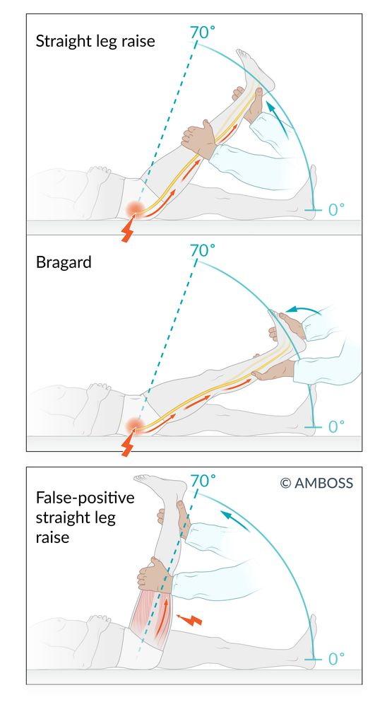
- Straight leg raise test (Lasègue test; top): With the patient supine and the knee extended, the examiner lifts the patient’s leg. The test is considered positive if lifting the leg to approximately 30–70° elicits radicular pain (i.e., pain that radiates from the buttock down the raised leg to below the knee). A positive test suggests lumbosacral nerve root impingement.
- Bragard test (middle): After a positive straight leg raise test, the leg is lowered (just enough for the pain to disappear), and the ankle is dorsiflexed. If dorsiflexion reproduces radiating pain, the Bragard test is considered positive, supporting the diagnosis of lumbosacral nerve root impingement.
- False-positive straight leg raise (bottom): If the straight leg raise test produces an aching pain confined to the posterior thigh (i.e., no radiating pain below the knee), typically when the leg is flexed to > 70°, it is considered a false-positive straight leg raise. This result suggests hamstring tightness or another musculoskeletal cause.
Diagnostics
- Initial / Screening:
- Clinical diagnosis in the absence of “red flag” symptoms (e.g., constitutional symptoms, nocturnal pain, progressive neuro deficits, trauma, history of cancer, IVDU).
- No routine imaging indicated for uncomplicated LBP < 6 weeks duration. c
- Imaging:
- Plain Radiographs (X-ray):
- Indicated if red flags present or initial conservative management fails.
- Findings: Loss of disk space height, osteophyte formation (syndesmophytes), endplate sclerosis, vacuum disk phenomenon (intradiskal gas).
- Magnetic Resonance Imaging (MRI):
- Confirmatory / Gold Standard for defining structural anatomy and pathology.
- Indicated for: Progressive/severe neurologic deficits, suspected infection/malignancy, or failure of conservative therapy > 4–6 weeks when invasive intervention is considered.
- Findings: Disk desiccation (“dark disk” on T2-weighted images), annular fissures (high-intensity zones), Modic endplate changes, herniations.
- Plain Radiographs (X-ray):
- Key Labs:
- ESR/CRP: Normal (used to rule out spondylodiscitis, epidural abscess, or malignancy).
Treatment
- First-line (Conservative Therapy - 4–6 weeks):
- Education & Activity: Maintain normal daily activities and light ambulation; avoid bed rest.
- Pharmacotherapy:
- NSAIDs (e.g., naproxen, ibuprofen) or acetaminophen first-line. c
- Short-term muscle relaxants (e.g., cyclobenzaprine) for acute painful paraspinal spasms (avoid long-term use).
- Avoid opioids for chronic non-cancer back pain.
- Physical Therapy (PT): Core stability, trunk strengthening, and McKenzie extension exercises once acute pain subsides.
- Second-line (Persistent Symptoms > 6 weeks):
- Re-evaluate with MRI.
- Fluoroscopy-guided epidural steroid injections (ESI) or facet joint injections (primarily beneficial if concurrent radicular irritation or facet arthropathy is present).
- Neuropathic adjuncts (e.g., duloxetine, gabapentin) if centralized chronic pain features develop.
- Refractory / Surgical Intervention:
- Indicated for persistent, disabling pain failing > 6–12 months of structured nonoperative management, or new progressive neurologic deficits.
- Options: Lumbar interbody fusion (arthrodesis) or total disk replacement (arthroplasty) in carefully selected candidates.