Shoulder (Glenohumeral) Dislocation
- Epidemiology & Mechanism:
- Anterior (~95%): Blow to arm in abduction, external rotation, and extension; or FOOSH.
- Posterior (2–4%): Seizures, electrocution/lightning, or direct anterior shoulder trauma.
- Inferior (<1%): Hyperabduction force (Luxatio erecta; arm locked overhead).
- Clinical Features:
- Anterior: Arm held in abduction & external rotation; “squared-off” shoulder with prominent acromion.
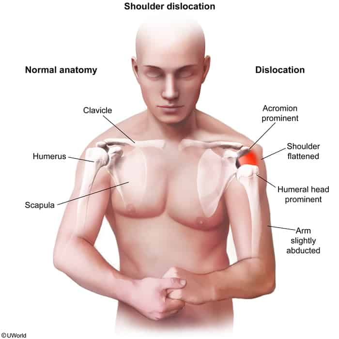
- Posterior: Arm locked in adduction & internal rotation; prominent coracoid, flattened anterior shoulder.
- NV Exam: Assess axillary nerve (lateral deltoid/“regimental badge” sensation & motor) and distal pulses before and after reduction. c
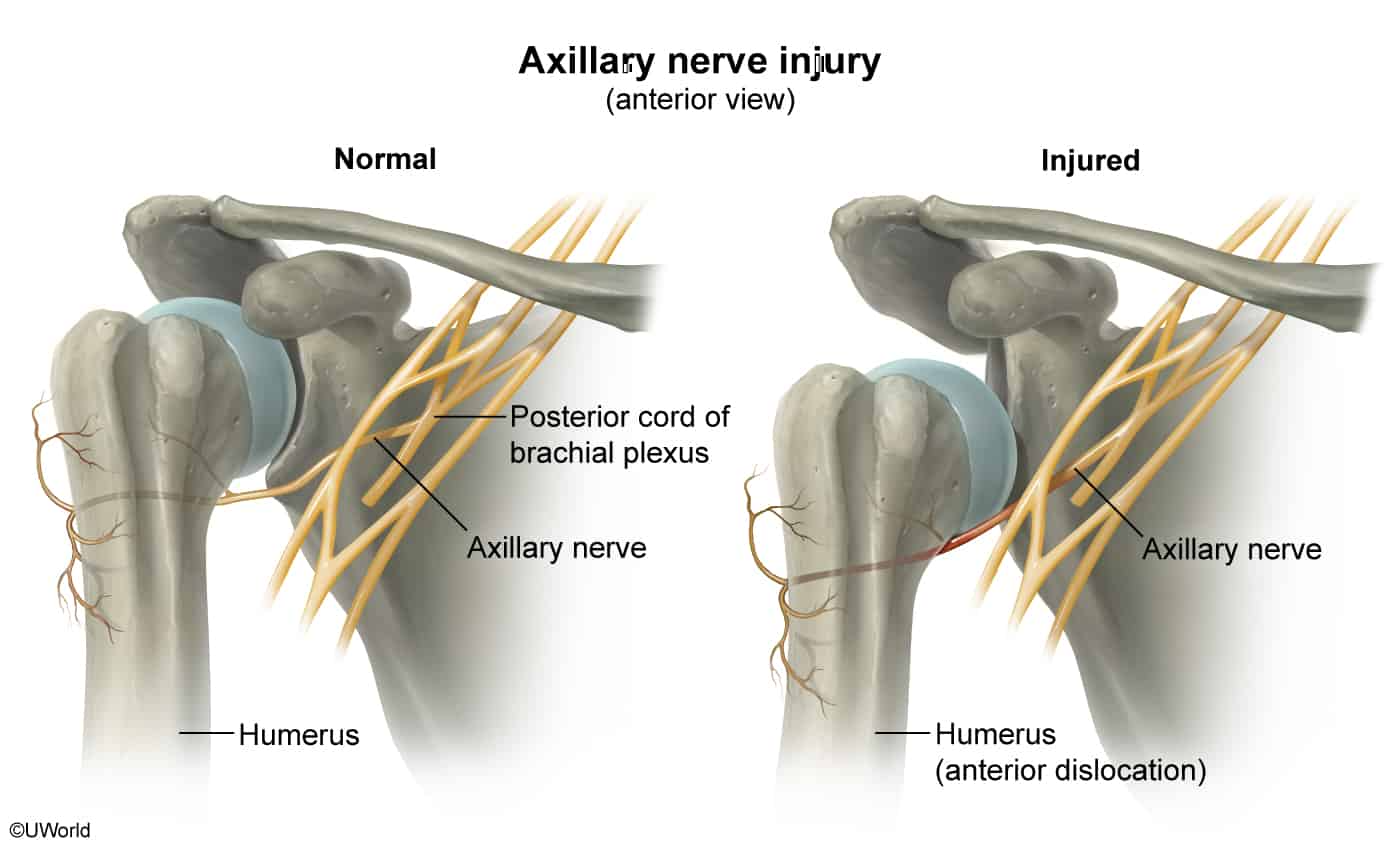
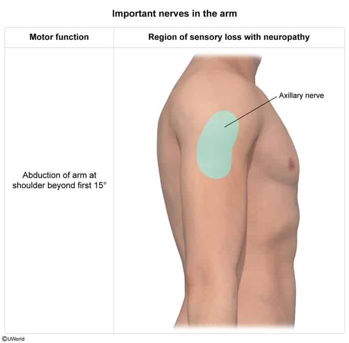
- Anterior: Arm held in abduction & external rotation; “squared-off” shoulder with prominent acromion.
- Diagnosis:
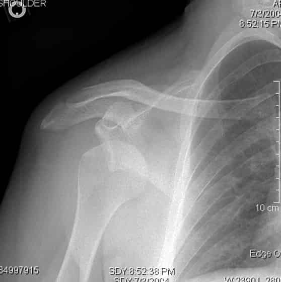
- Best Initial Test: XR shoulder trauma series (AP, Scapular ‘Y’, and Axillary lateral view [mandatory to rule out posterior dislocation]).
- Anterior Lesions:
- Bankart lesion: Tear of anterior-inferior glenoid labrum ± bony fracture.
- Hill-Sachs lesion: Compression fracture of posterolateral humeral head.
- Posterior Lesions:
- “Lightbulb sign” on AP view (fixed internal rotation).
- Reverse Hill-Sachs lesion: Anteromedial humeral head impaction.
- Best Initial Test: XR shoulder trauma series (AP, Scapular ‘Y’, and Axillary lateral view [mandatory to rule out posterior dislocation]).
- Management Hierarchy:
- Pre-reduction NV exam: Mandatory documentation of axillary nerve and vascular status.
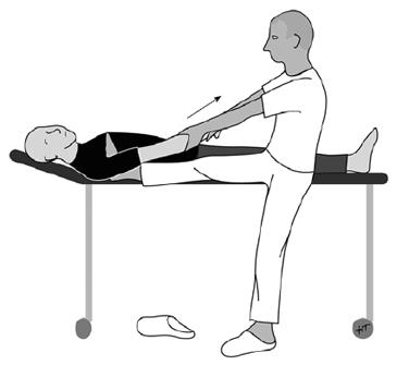
- Closed reduction: Emergent reduction under procedural sedation or intra-articular lidocaine (e.g., traction-countertraction).
- Post-reduction NV exam & repeat XR: Confirm joint relocation and rule out iatrogenic fracture.
- Immobilization & Rehab:
- Young pts: Sling for 1–3 weeks followed by PT.
- Elderly pts (>40 yo): Brief sling use (<1 week) to prevent adhesive capsulitis.
- Surgery (Open reduction / Labral repair): Indicated for irreducible dislocation, open injury, neurovascular entrapment, or recurrent instability.
Mnemonic
拍拍肩膀说:嗨H 拍拍屁股说:啊A
Hip Dislocation
- Patho/Etiology
- Requires significant force; often from motor vehicle accidents (dashboard injury) or falls from a height.
- Posterior (>90%): Femoral head is forced posteriorly out of the acetabulum.
- Anterior (<10%): Usually from forced abduction.
- Clinical Presentation
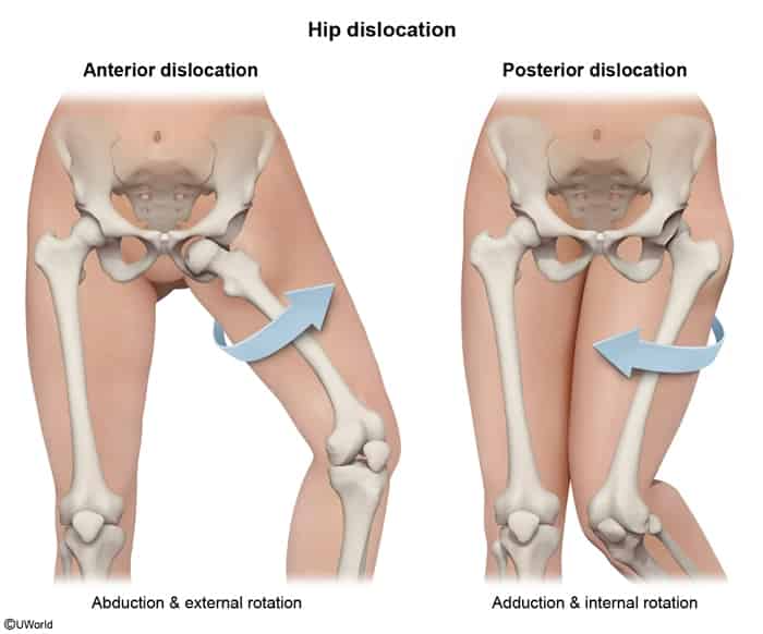
- Posterior: The affected limb is shortened, adducted, and internally rotated. This is the classic, high-yield presentation.
- Anterior: Limb is abducted and externally rotated.
- Diagnosis
- X-ray (AP pelvis): Confirms diagnosis. In posterior dislocation, the femoral head appears smaller than the contralateral side. A CT scan is often done post-reduction to check for associated fractures.
- Management
- Orthopedic emergency.
- Prompt closed reduction under sedation to decrease the risk of avascular necrosis (Allis technique).
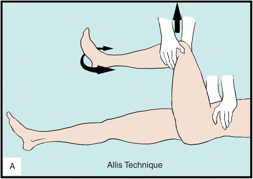
- Open reduction if closed reduction fails or if there are associated fractures.
- Key Complications
- Avascular necrosis (AVN) of the femoral head: The most serious complication; risk increases with time to reduction.
- Sciatic nerve injury: Common with posterior dislocations, presenting with foot drop (impaired dorsiflexion).
- Post-traumatic arthritis: The most common long-term complication.
Elbow Dislocation
- Patho/Etiology
- Second most common major joint dislocation in adults; most common in children.
- Usually caused by a fall on an outstretched hand (FOOSH) with the elbow in extension.
- Posterior dislocation is the most common type.
- Clinical Presentation
- Presents with severe pain, swelling, and inability to bend the elbow. The olecranon is prominent posteriorly.
- Diagnosis
- X-ray (AP and lateral): Confirms dislocation. Important to assess for associated fractures.
- “Terrible triad” of the elbow: A complex dislocation involving a posterior dislocation, a radial head fracture, and a coronoid process fracture.
- Management
- Immediate closed reduction after a careful neurovascular exam.
- Post-reduction, the elbow is splinted in flexion (around 90 degrees). Early range of motion is crucial to prevent stiffness.
- Key Complications
- Neurovascular injury:
- Brachial artery injury: Can be limb-threatening. Always check for distal pulses before and after reduction.
- Median and ulnar nerve injuries are also common.
- Compartment syndrome: A risk, especially with associated fractures.
- Contracture/Stiffness: Loss of terminal extension is a very common long-term complication.
- Neurovascular injury:
Knee (Tibiofemoral) Dislocation
- Patho/Etiology
- A rare but limb-threatening orthopedic emergency, often from high-energy trauma (MVA, falls).
- Can occur with lower energy in morbidly obese individuals.
- Up to 50% of knee dislocations spontaneously reduce before evaluation, so a high index of suspicion is required.
- Clinical Presentation
- Gross deformity and instability of the knee. If reduced, may present with significant swelling and multi-ligamentous instability.
- Diagnosis
- Based on clinical suspicion, even if the joint is reduced.
- Ankle-brachial index (ABI): A crucial screening tool. An ABI <0.9 warrants further vascular imaging.
- CT angiography: To rule out vascular injury.
- Management
- Immediate reduction. Do not delay for imaging if there are signs of vascular compromise.
- Careful and repeated neurovascular checks are critical.
- Admission for observation and serial exams is standard.
- Key Complications
- Popliteal artery injury (up to 40%): High risk of amputation if not recognized and treated promptly (within 8 hours). This is the most feared complication.
- Common peroneal (fibular) nerve injury: Presents with foot drop and numbness on the dorsum of the foot.
- Multiple ligament tears (ACL, PCL, etc.): Nearly always present and require surgical reconstruction.