Common lower extremity injury; high incidence in young athletic males and older osteoporotic females.
Risk Factors:
Low-energy rotational or twisting trauma (inversion/supination or eversion/pronation).
High-energy axial load (e.g., MVC, fall from height).
Osteoporosis / osteopenia.
Diabetic peripheral neuropathy (predisposes to severe displacement and Charcot neuroarthropathy).
Clinical Features
Symptoms: Immediate severe pain, rapid swelling, ecchymosis, deformity, inability to bear weight.
Physical Exam:
Bony tenderness over medial malleolus, lateral malleolus, and/or posterior malleolus.
Neurovascular (NV) status: Dorsalis pedis and posterior tibial pulses, capillary refill, sensation in deep peroneal and tibial nerve distributions.
Skin evaluation: Assess for tenting (risk of ischemic necrosis) and open wounds.
Palpation of proximal fibula is mandatory to rule out high fibular fracture (Maisonneuve fracture).
Diagnosis
Initial / Decision Rule:
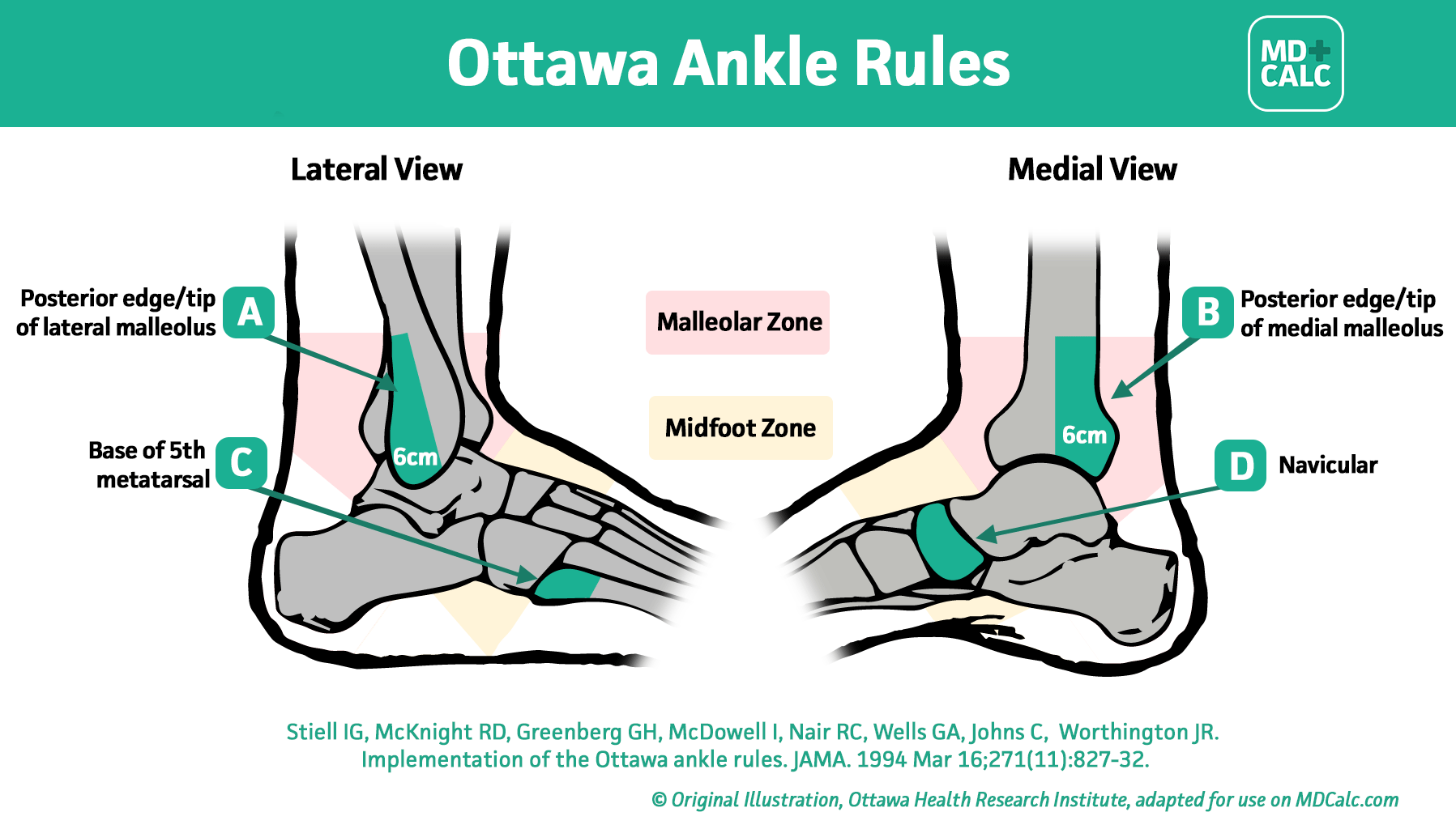
Ottawa Ankle Rules (determines need for imaging; sensitivity ~99%): c
Plain radiographs indicated if pain in the malleolar zone AND:
Bone tenderness along the posterior edge or tip of the lateral malleolus (distal 6 cm), OR
Bone tenderness along the posterior edge or tip of the medial malleolus (distal 6 cm), OR
Inability to bear weight both immediately after injury and in the ED (4 steps).
Imaging:
Initial / Gold Standard for Basic Fracture: 3-view Plain Ankle Radiographs (AP, Lateral, and Mortise view [AP in 15–20° internal rotation]).
Mortise view: Evaluates the medial clear space; widening > 4–5 mm indicates deltoid ligament disruption and syndesmotic instability.
Full-length Tibia/Fibula Radiographs: Indicated if medial malleolar tenderness/widening occurs without a clear distal fibular fracture on ankle XR (rules out Maisonneuve fracture).
CT Ankle: Non-contrast CT for complex intra-articular patterns (e.g., triplane, Tillaux, pilon fractures) and pre-operative planning.
Diff by maximal tenderness localized over ligaments (anterior talofibular ligament [ATFL], calcaneofibular ligament) rather than bony malleoli; Ottawa rules negative; normal radiographs.
Maisonneuve Fracture:
Diff by spiral fracture of the proximal third of the fibula associated with disruption of the distal tibiofibular syndesmosis and deltoid ligament rupture or medial malleolus fracture. Must palpate proximal fibula in all ankle injuries.
Achilles Tendon Rupture:
Diff by sudden “pop” in posterior ankle/calf, palpable tendon gap, and positive Thompson test (absence of plantar flexion upon calf squeeze); ankle mortise intact on XR.
Fifth Metatarsal Base Fracture (Jones vs. Pseudo-Jones):
Diff by tenderness localized over the tuberosity or diaphysis of the fifth metatarsal; foot XR diagnostic.
Pilon Fracture (Tibial Plafond):
Diff by high-energy axial loading mechanism; extensive comminution and impaction of the distal tibial articular surface visible on XR/CT.
Management
Immediate Emergency / Stabilization:
Closed Reduction: Perform urgently if severe deformity, skin tenting, or NV compromise is present; do not delay for imaging if critical limb ischemia exists.
Open Fracture: Immediate IV antibiotics (e.g., Cefazolin ± Aminoglycoside), tetanus prophylaxis, sterile dressing, and emergent surgical debridement/irrigation.
Temporary Immobilization: Well-padded posterior splint with U-splint (stirrup) in neutral position; Non-Weight Bearing (NWB); elevate limb and apply ice to mitigate soft tissue swelling.
Definitive Management:
Conservative / First-line for Stable Fractures:
Isolated, non-displaced Weber A fractures or stable Weber B fractures without medial clear space widening.
Short-leg cast or rigid walking boot for 4–6 weeks with protected weight-bearing.
Surgical / First-line for Unstable Fractures:
Indications: Displaced bimalleolar, trimalleolar, Weber C, unstable Weber B (medial clear space > 4 mm), or open fractures.
Open Reduction and Internal Fixation (ORIF): Rigid plate and screw fixation of fibula/tibia; syndesmotic screw fixation if syndesmosis remains widened.
Second-line / Refractory:
External Fixation (Span-Scan-Plan): Temporary bridging external fixator used when severe soft tissue swelling, blister formation, or open contamination precludes immediate internal fixation; delayed definitive ORIF performed once soft tissues wrinkle (typically 10–14 days).
Complications
Post-Traumatic Osteoarthritis: Most frequent long-term complication; risk increases with residual intra-articular incongruity > 1 mm.
Compartment Syndrome: Rare in isolated low-energy ankle fractures, but risk rises with high-energy crush or pilon variants.
Wound Dehiscence / Infection: High incidence in poorly controlled DM, peripheral vascular disease, active smokers, or when surgery is performed through acutely swollen/compromised soft tissue.
Malunion / Nonunion: Leads to persistent ankle instability, chronic pain, and early joint destruction.
Complex Regional Pain Syndrome (CRPS): Characterized by burning pain, allodynia, sudomotor, and vasomotor changes post-injury/surgery.