Normal Physiologic Alignment & Progression
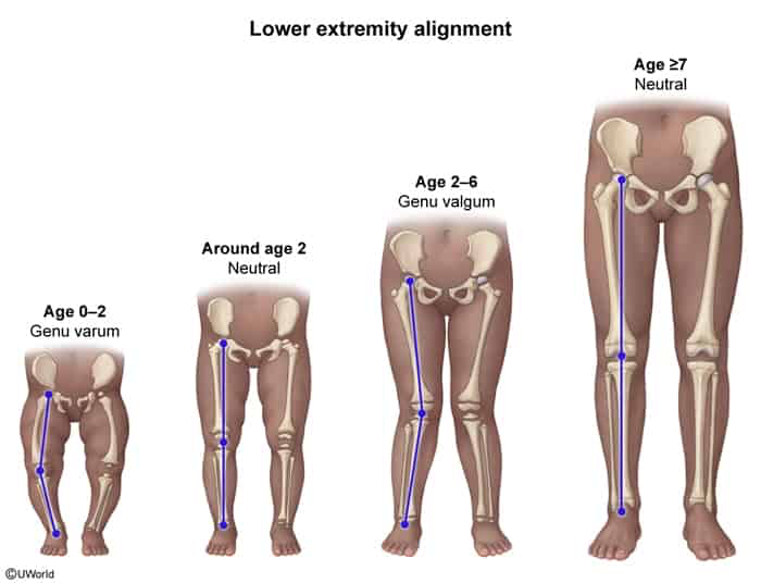
- Birth to 18–24 months: Physiologic genu varum (bowlegs).
- Symmetric bowing.
- Max bowing occurs at ~6–12 months.
- Normalizes to neutral alignment by 18–24 months.
- 2 to 5 years: Physiologic genu valgum (knock-knees).
- Alignment shifts to valgus; peaks at ~3–4 years of age.
- 6 to 7 years to adulthood: Gradually resolves to normal adult alignment (~5–7° of valgus).
- Unilateral or asymmetric presentation.
- Severe or progressive angulation outside the typical age range (e.g., genu varum worsening after age 2; genu valgum persisting after age 7).
- Associated short stature (<5th percentile for age).
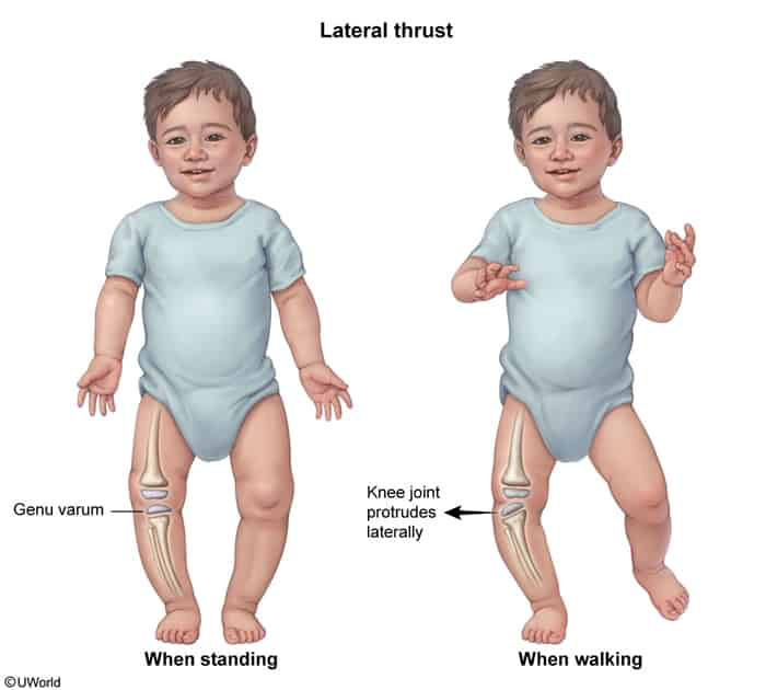
- Pain, limp, or abnormal gait (e.g., lateral thrust during stance phase).
- Intercondylar distance (femoral) > 6 cm in genu varum.
- Intermalleolar distance > 8 cm in genu valgum.
- Concomitant systemic manifestations (e.g., craniotabes, rachitic rosary, dental abnormalities).
Genu Varum (Bowlegs)
Etiologies
- Physiologic Genu Varum:
- Bilateral, symmetric, painless; normal stature; self-corrects by age 2.
- Blount Disease (Tibia Vara):
- Pathologic disruption of the posteromedial proximal tibial physis.
- Risk Factors: African descent, early walking (<1 year), childhood obesity.
- Types:
- Infantile (1–3 years): Often bilateral, severe progressive varus, medial tibial torsion.
- Adolescent (>8 years): Unilateral, associated w/ rapid weight gain.
- Imaging: Sharp, medial beaking of proximal tibial metaphysis, sloped epiphysis.
- Nutritional Rickets:
- Defective mineralization of osteoid secondary to Vitamin D deficiency.
- Signs: Craniotabes, rachitic rosary (costochondral swelling), widened wrists, bowing.
- Labs: ↓ / normal Ca, ↓↓ Phos, ↑↑ ALP, ↑ PTH, ↓ 25-OH Vit D.
- Imaging: Metaphyseal fraying, cupping, and widening.
- Skeletal Dysplasias:
- E.g., Achondroplasia (FGFR3 mutation: rhizomelic limb shortening, macrocephaly, frontal bossing).
- Post-Traumatic / Post-Infectious:
- Physeal injury (Salter-Harris fractures) or osteomyelitis leading to asymmetric growth arrest.
Genu Valgum (Knock-Knees)
Etiologies
- Physiologic Genu Valgum:
- Symmetric, age 2–5, resolves spontaneously by age 7–8; intermalleolar distance < 8 cm.
- Renal Osteodystrophy (“Renal Rickets”):
- Chronic kidney disease (CKD) → impaired 1-alpha-hydroxylase → ↓ 1,25-(OH)2 Vit D + phosphate retention → secondary hyperparathyroidism.
- Common cause of severe pathologic genu valgum in older children/adolescents.
- Post-Traumatic (Cozen Phenomenon):
- Valgus deformity following a proximal tibial metaphyseal fracture (transient asymmetric hypervascularity and overgrowth of medial physis).
- Skeletal Dysplasias & Connective Tissue Disorders:
- E.g., Ehlers-Danlos syndrome, Marfan syndrome, Morquio syndrome.
Diagnostic Workup
Initial / Physical Exam
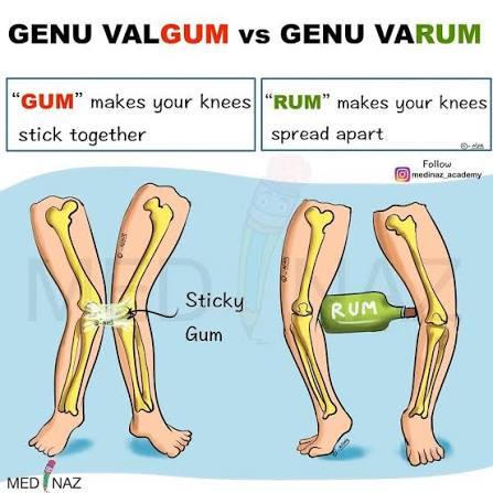
- Measure Intercondylar Distance (varum: knees apart with ankles touching).
- Measure Intermalleolar Distance (valgum: ankles apart with knees touching).
- Evaluate rotational profile: Assess for internal/external tibial torsion and femoral anteversion.
- Gait analysis: Assess for lateral thrust (knee shifts laterally during stance phase → indicates Blount disease or ligamentous laxity).
Imaging
- Full-Length Standing AP & Lateral Radiographs (hips to ankles, weight-bearing):
- Indicated only if red flags are present (asymmetry, pain, age > 2 for varum, severe progression).
- Measures mechanical axis deviation (MAD) and tibial-femoral angle.
- Identifies metaphyseal beaking (Blount disease) or cupping/fraying (Rickets).
Laboratory Evaluation
- Indicated if rickets, renal disease, or skeletal dysplasia suspected:
- Serum Ca, Phos, ALP, Cr, BUN.
- 25-OH Vit D (screen for nutritional deficiency).
- 1,25-(OH)2 Vit D (if CKD or Vit D-dependent rickets suspected).
- Intact PTH.
Differential Diagnostics
- Physiologic Genu Varum vs. Blount Disease:
- Physiologic: Resolves by 18–24 mo; diffuse bowing of femur and tibia; normal tibial metaphysis.
- Blount: Progressive; localized to proximal medial tibia; medial metaphyseal beaking and stepping on XR.
- Blount Disease vs. Nutritional Rickets:
- Blount: Normal serum labs (Ca, Phos, ALP normal); isolated tibial physis deformity.
- Rickets: Abnormal labs (↑ ALP, ↓ Phos); generalized skeletal involvement (widened wrists, rachitic rosary, diffuse fraying).
- Physiologic Genu Valgum vs. Renal Osteodystrophy:
- Physiologic: Peak age 3–4, normal labs, no systemic signs.
- Renal Osteodystrophy: Older child/adolescent, azotemia (↑ Cr/BUN), ↑ Phos, ↓ Ca, ↑ PTH.
Management
Physiologic Varum / Valgum
- Reassurance and Observation:
- No orthotics, special shoes, or braces required (ineffective and unnecessary).
- Follow-up clinically every 4–6 months to document expected resolution.
Pathologic Genu Varum
- Infantile Blount Disease:
- Initial: KAFO (Knee-Ankle-Foot Orthosis) brace if age < 3 years and early stage (Langenskiöld stage I–II).
- Surgical: Proximal tibial realignment osteotomy if no response to bracing by age 4 or stage ≥ III.
- Adolescent Blount Disease:
- Bracing is ineffective.
- Surgical: Guided growth (temporary hemiepiphysiodesis using 8-plates) if sufficient growth remains; corrective osteotomy if near skeletal maturity.
- Nutritional Rickets:
- Medical: High-dose oral Vitamin D (Ergocalciferol/Cholecalciferol) + supplemental calcium.
- Skeletal deformity corrects spontaneously once remineralization occurs; surgery reserved for persistent deformities after metabolic normalization.
Pathologic Genu Valgum
- Mild to Moderate (Growing Child):
- Guided growth (medial hemiepiphysiodesis) to allow lateral side to catch up.
- Severe / Skeletally Mature:
- Corrective femoral/tibial osteotomy.
- Renal Osteodystrophy:
- Phosphate binders, Calcitriol, dietary phosphate restriction; optimize dialysis/renal transplant.
High-Yield USMLE Pearls
- Timeline Rule: Bowlegs up to age 2, knock-knees from age 2 to 5, normal adult alignment by age 7.
- Lateral thrust during gait is a pathognomonic clinical clue for Blount disease or severe ligamentous instability.
- Never order radiographs or braces for an asymptomatic, normally growing 14-month-old with mild, symmetric bowlegs → select Reassurance/Observation.
- Widened wrist physes + metaphyseal fraying/cupping on XR = Rickets; isolated proximal medial tibial beaking in an obese, early-walking toddler = Blount disease.