Palpable gap/defect in the tendon (usually 2–6 cm proximal to calcaneal insertion).
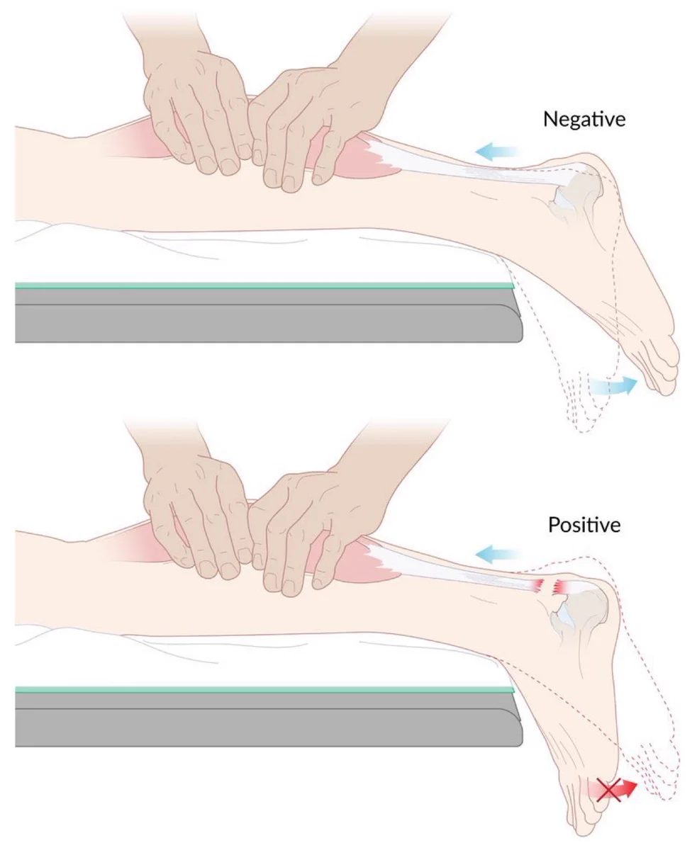
Positive Thompson test (Simmonds test): Pt prone with feet hanging off the table; squeezing the calf produces absent or diminished passive plantarflexion. c
Inability to perform single-leg heel raise.
Weakness in active plantarflexion (weak compensatory plantarflexion may remain via posterior tibialis, peroneals, and toe flexors).
Diagnosis
Clinical Diagnosis: Primarily diagnosed by characteristic Hx and (+) Thompson test with a palpable gap.
Initial / Bedside Imaging:
Ultrasound (US): High sensitivity/specificity; confirms complete vs. partial tear, tendon retraction, and hematoma.
Confirmatory / Gold Standard Imaging:
MRI ankle: Best modality; assesses tear extent, chronicity, tendon retraction gap, and soft tissue status for surgical planning (indicated when clinical diagnosis is equivocal).
Plain Radiographs (X-ray):
Normal or soft-tissue swelling; rule out associated calcaneal avulsion fracture.
Differential Diagnostics
Achilles Tendinopathy / Tendinitis:
Gradual onset morning stiffness/pain; tenderness along tendon; (-) Thompson test, no palpable defect.
Gastrocnemius Tear (“Tennis Leg”):
Pain localized to the medial proximal calf belly (musculotendinous junction); intact Achilles tendon; (-) Thompson test.
Ankle Sprain (ATFL / CFL Tear):
Inversion injury mechanism; tenderness over the lateral malleolus/ligaments; intact calf squeeze and heel raise.
Calcaneal Stress Fracture:
Gradual onset heel pain in runners/military recruits; pain on lateral-to-medial calcaneal compression (calcaneal squeeze test).
Retrocalcaneal Bursitis:
Pain anterior to the Achilles tendon insertion; pain on deep palpation anterior to tendon; intact plantarflexion mechanics.
Management
Acute / Initial Stabilization:
RICE (Rest, Ice, Compression, Elevation) + non-weight-bearing with crutches.
Immobilization: Rigid splinting in slight plantarflexion (equinus position) to approximate tendon ends.
Analgesia (NSAIDs, acetaminophen).
Definitive Treatment:
Non-operative Management:
Indications: Low physical demand, elderly, sedentary pts, or high surgical/anesthetic risk (e.g., severe DM, PVD).
Regimen: Functional bracing or serial casting in equinus, gradually moving toward neutral over 8–12 wks + early functional rehab.
Operative Repair:
Indications: Young, athletic, active pts, delayed presentation (> 1–2 wks), or re-rupture.
Outcome: Lower re-rupture rate compared to conservative therapy, but higher risk of wound complications.
Refractory / Chronic Tears (> 4–6 weeks delay):
Open reconstruction with tendon transfer (e.g., flexor hallucis longus transfer) or V-Y tendinoplasty.
Complications
Tendon Re-rupture (higher rate in non-operative vs. operative management).
Surgical Complications:
Wound dehiscence, skin necrosis, superficial/deep surgical site infection.
Sural nerve injury / neuropraxia (leads to lateral foot numbness).
Deep Vein Thrombosis (DVT) / PE secondary to lower extremity immobilization.