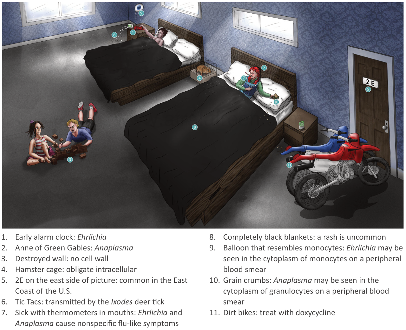
Ehrlichiosis
- Pathogens
- Vectors
- Lone star tick (Amblyomma americanum): E. chaffeensis and E. ewingii
- Deer tick (Ixodes scapularis): E. muris eauclairensis
- Distribution
- Mainly east of the Rocky Mountains
- Also some cases in the Southwest
- Clinical features
- Systemic: Sudden high fever, chills, severe frontal headache, myalgias, malaise.
- GI: Nausea, vomiting, diarrhea, abdominal pain.
- Rash: Uncommon in adults (<30%), more common in children (~60%). Maculopapular or petechial, usually sparing the face.
- CNS: Altered mental status, confusion, stiff neck, seizures (indicates severe infection/meningoencephalitis).
- Diagnostics
- Key Labs (High-Yield Triad):
- Leukopenia (specifically lymphopenia and neutropenia).
- Thrombocytopenia.
- Elevated transaminases (AST/ALT) and LDH.
- Initial/Screening:
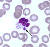
- Peripheral blood smear: Morulae inside monocytes (mulberry-like intracytoplasmic inclusions; low sensitivity, high specificity).
- Confirmatory:
- Whole-blood PCR (most sensitive test in acute phase before seroconversion).
- Serology (IFA): 4-fold rise in IgG antibody titers between acute and convalescent phases.
Tick paralysis
- Pathophysiology: Salivary neurotoxin impairs presynaptic acetylcholine (ACh) release at NMJ after prolonged tick feeding (Dermacentor andersoni/variabilis).
- Clinical Presentation: c
- Rapidly ascending flaccid paralysis (progresses over hours to days).
- Areflexia/hyporeflexia, gait ataxia, afebrile.
- Normal sensation and normal autonomic function.
- Late signs: Bulbar weakness (dysphagia, dysarthria) and diaphragmatic paralysis/respiratory failure.
- Diagnosis:
- Initial/Best: Meticulous full-body skin exam (especially scalp, hairline, and skin folds).
- Lumbar Puncture: Normal CSF (rules out Guillain-Barré syndrome; no albuminocytologic dissociation).
- High-Yield Differentials:
- Guillain-Barré Syndrome (GBS): Slower onset (days–weeks), antecedent URI/GI infection, autonomic instability, ↑ CSF protein with normal WBC. c
- Botulism: Descending paralysis, early cranial nerve deficits (pupillary dilation, ptosis, diplopia), autonomic dysfunction.
- Spinal Cord Compression: Distinct sensory level, hyperreflexia (late), bowel/bladder dysfunction.
- Management:
- Definitive: Complete mechanical tick removal (rapid clinical recovery within 24–48 hours).
- Supportive: Monitor respiratory status (FVC / NIF); intubate if signs of impending respiratory failure.
- Note: No antivenom or antibiotics indicated for North American tick paralysis.
Tularemia
- Epidemiology & Risk Factors
- Organism: Francisella tularensis (Gram-negative intracellular coccobacillus).
- Exposures: Rabbit skinning/hunting, tick/deer fly bites, lawn mowing over carcasses (endemic: Martha’s Vineyard, AR, MO, OK).
- Bioterrorism: Category A agent (aerosolized risk).
- Clinical Presentation
- Ulceroglandular (most common): Painful punched-out skin ulcer + tender regional lymphadenopathy. c
- Pneumonic: Inhalation of aerosolized bacteria → high fever, dyspnea, hilar adenopathy (high mortality).
- Other forms: Glandular (no ulcer), Oculoglandular (conjunctivitis + preauricular LN), Oropharyngeal (ingestion).
- Diagnosis
- Best Initial/Confirmatory: Serology (4-fold titer rise) or PCR.
- Contraindicated: Bacterial culture (high hazard for laboratory-acquired infection; requires BSL-3).
- Differential Diagnosis
- Bartonella henselae (Cat scratch): Subacute/indolent, painless papule.
- Bacillus anthracis (Cutaneous anthrax): Painless black eschar + marked edema.
- Sporothrix schenckii: Painless ascending nodular lymphangitis (rose gardener).
- Management
- Severe / Hospitalized: IV Gentamicin or Streptomycin.
- Mild / Outpatient: PO Doxycycline or Ciprofloxacin (14–21 days).
- Post-Exposure Prophylaxis: PO Doxycycline or Ciprofloxacin x 14 days.