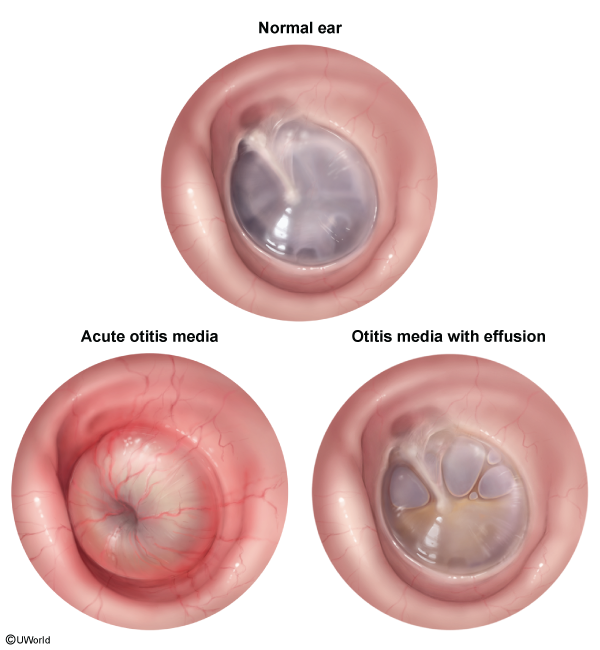
Most common cause of pediatric hearing impairment.
Peak incidence: Ages 6 months to 4 years.
Pathophysiology: Fluid accumulation in middle ear cleft without acute systemic/local signs of infection; often follows resolution of Acute Otitis Media (AOM) or arises from Eustachian tube dysfunction (ETD).
Risk Factors:
Recent episode of AOM (fluid persists in 70% at 2 wks, 40% at 1 mo, 10-25% at 3 mos).
TM appearance: Amber, dull, or gray; translucent or opaque.
No significant bulging (may be neutral or retracted).
Air-fluid levels or clear fluid bubbles visible behind the TM.
Confirmatory / Objective Testing:
Tympanometry: Type B tracing (flat curve indicating decreased TM compliance and middle ear fluid) or Type C (negative pressure).
Audiometry (Formal Hearing Test): Demonstrates Conductive Hearing Loss (CHL); indicated if effusion persists ≥ 3 months or if developmental delays are suspected.
Differential Diagnostics
Acute Otitis Media (AOM):
Diff: Presents with acute signs of inflammation (fever, marked TM bulging, erythema, severe otalgia). OME lacks acute inflammatory signs and significant bulging.
Eustachian Tube Dysfunction (ETD without effusion):
Diff: TM is retracted with prominent bony landmarks, but lacks air-fluid levels, bubbles, or middle ear fluid on pneumatic otoscopy/tympanometry.
Bullous Myringitis:
Diff: Presence of painful, fluid-filled vesicles/bullae directly on the outer surface of the TM; severe acute otalgia.
Otitis Externa:
Diff: Pain elicited with manipulation of the pinna or tragus; canal erythema and edema; TM is typically clear and mobile.
Cholesteatoma:
Diff: Chronic drainage, white keratinaceous debris or mass in retraction pocket / attic region of TM; progressive bone erosion.
Management
First-Line / Initial:
Watchful waiting and observation: Re-examine in 3 months.
Most cases resolve spontaneously within 3 months. c
Do NOT give: Systemic/topical antibiotics, oral/nasal steroids, antihistamines, or decongestants (no clinical benefit in OME).
Second-Line / Surgical Intervention:
Indications:
Bilateral OME persistent for ≥ 3 months with documented hearing loss / speech delay.
Recurrent/chronic OME with structural TM damage (e.g., severe retraction, atelectasis).
High-risk children (e.g., Down syndrome, cleft palate, sensorineural hearing loss, autism).
Procedure of Choice: Tympanostomy tube insertion (myringotomy with ventilation tubes).
Adjunctive: Adenoidectomy if concurrent chronic nasal obstruction or recurrent adenoiditis in children ≥ 4 years old.
Complications
Conductive Hearing Loss (CHL) (can fluctuate).
Delayed speech and language development.
TM structural abnormalities:
Tympanosclerosis (calcium plaques).
Adhesive otitis / TM atelectasis.
Retraction pockets predisposing to cholesteatoma formation.