Epidemiology & Risk Factors
- Pathogens: Opportunistic fungi of order Mucorales (Rhizopus, Mucor, Lichtheimia).
- Primary Risk Factor: Uncontrolled DM with DKA (high glucose and acidic environment allow fungal proliferation via ketone reductase).
- Other Risk Factors:
- Iron overload or Deferoxamine therapy (fungus uses iron as growth factor).
- Hematologic malignancies (e.g., AML), prolonged neutropenia.
- Solid organ or hematopoietic stem cell transplant.
- High-dose chronic systemic corticosteroid use.
Clinical Features
- Rhinocerebral Mucormycosis (classic presentation in DKA):
- Rapidly progressive facial pain, headache, fever, purulent nasal discharge.
- Black necrotic eschar on nasal turbinates, septum, or hard palate (pathognomonic, secondary to tissue ischemia/angioinvasion).
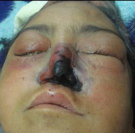
- Periorbital swelling, proptosis, ophthalmoplegia, loss of vision (orbital apex syndrome).
- Cranial nerve deficits (CN III, IV, VI, V1, V2).
- Altered mental status, lethargy (CNS extension).
- Pulmonary Mucormycosis (common in severe neutropenia):
- Fever, cough, hemoptysis, pleuritic chest pain.
- Cutaneous Mucormycosis:
- Necrotic skin lesions in trauma or burn patients.
Diagnosis
- Initial: High clinical suspicion in at-risk patient (DKA + facial pain/black eschar) →\rightarrow→ direct visualization via nasal endoscopy.
- Imaging: CT or MRI of sinuses/brain (shows sinus opacification, bone destruction, retro-orbital extension, or carotid artery thrombosis).
- Confirmatory/Gold Standard: Tissue biopsy with histopathology.
- Histopathology: Broad, non-septate (or sparingly septate) hyphae branching at right angles (90°).
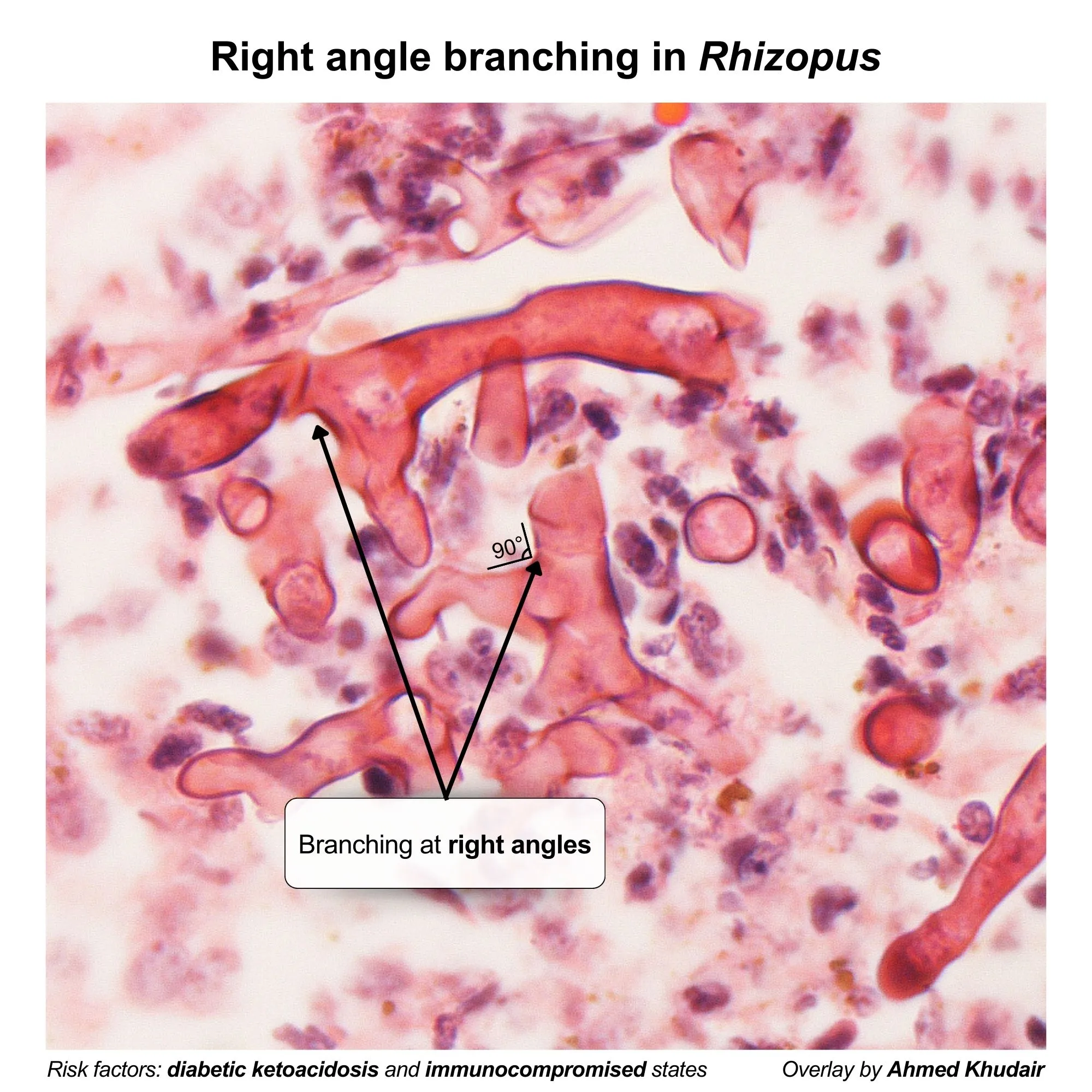
- Hallmark Feature: Angioinvasion leading to vascular thrombosis and tissue necrosis.
- Fungal culture (confirmative, but treatment must not be delayed for results).
Differential Diagnostics
- Invasive Aspergillosis:
- Differentiated by narrow, septate hyphae branching at acute angles (45°).
- More common in prolonged neutropenia/transplant; rare in DKA.
- Bacterial Orbital Cellulitis / Sinusitis:
- Absence of black necrotic eschar; no response to broad-spectrum Abx.
- Cavernous Sinus Thrombosis (Bacterial):
- Similar CN involvement, but lacks necrotic mucosal lesions and fungal hyphae on biopsy.
Management
- Immediate Surgical Debridement:
- Surgical emergency; radical debridement of all necrotic tissue is essential for survival.
- First-Line Antifungal:
- High-dose IV Liposomal Amphotericin B (start immediately upon clinical suspicion before biopsy confirmation).
- Correction of Underlying Etiology:
- Aggressive management of DKA (IV insulin, fluids).
- Discontinue iron chelators (deferoxamine).
- Taper immunosuppressive therapy/steroids if feasible.
- Step-Down / Maintenance Therapy:
- Oral Isavuconazole or Posaconazole after initial clinical response to amphotericin B.