Epidemiology & Risk Factors

  • Etiology: Echinococcus granulosus (cystic echinococcosis; most common) or E. multilocularis (alveolar echinococcosis).
  • Hosts:
    • Definitive host: Dogs/canines.
    • Intermediate host: Sheep, cattle, goats.
    • Accidental intermediate host: Humans (ingestion of food/water contaminated w/ dog feces containing eggs).
  • Risk Factors: Rural sheep-farming regions (Mediterranean, Middle East, South America, Central Asia, Australia).

Clinical Features

  • Latent for years/decades; usually asymptomatic until cyst grows large. c
  • Hepatic involvement (~60-70%, right lobe > left):
    • RUQ dull discomfort/pain, hepatomegaly, palpable mass, abdominal distension.
    • Biliary compression -> jaundice, pruritus.
  • Pulmonary involvement (~20-30%):
    • Cough, chest pain, hemoptysis, dyspnea.
  • Cyst rupture:
    • Fever, urticaria, severe anaphylactic shock, and rapid intraperitoneal dissemination.

Diagnosis

  • Initial Test: RUQ Ultrasound (shows simple/complex fluid-filled cyst with daughter cysts, internal septations, or “hydatid sand”).
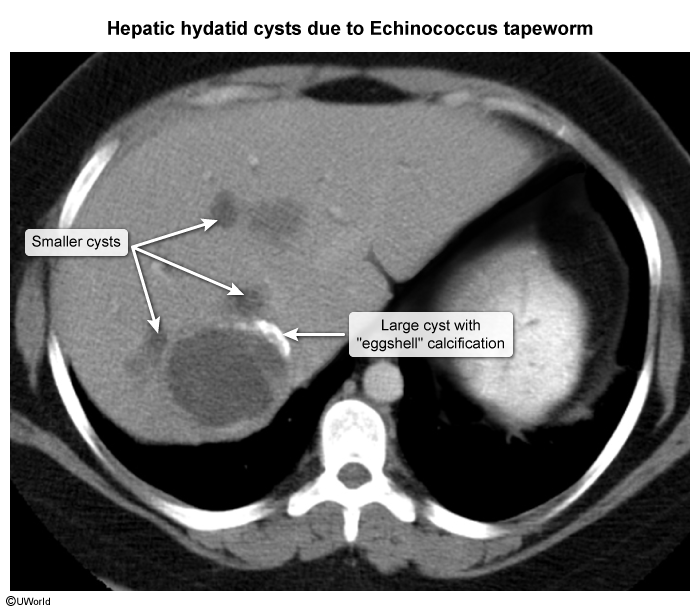
  • Confirmatory Imaging: Abdominal CT (demonstrates well-defined, unilocular or multilocular cyst with “eggshell” wall calcification).
  • Key Labs:
    • Eosinophilia (present in <50%, increased if cyst leaks).
    • IgG Serology (ELISA): Confirms diagnosis; high sensitivity/specificity.
  • Contraindication: Percutaneous diagnostic aspiration/biopsy is strictly CONTRAINDICATED due to high risk of leakage -> anaphylaxis and peritoneal seeding.

Differential Diagnostics

  • Amebic Liver Abscess (Entamoeba histolytica):
    • Diff: Acute onset w/ high fever, travel history, “anchovy paste” fluid, no calcifications or daughter cysts; treat w/ Metronidazole + Paromomycin.
  • Pyogenic Liver Abscess:
    • Diff: Sepsis signs (high fever, leukocytosis), history of cholangitis/diverticulitis, CT shows solitary/multiple hypodense ring-enhancing lesions; treat w/ Abx + drainage.
  • Simple Hepatic Cyst:
    • Diff: Congenital, completely asymptomatic, incidental finding, thin-walled on US, no calcifications/internal daughter cysts, normal serology.
  • Hepatic Hemangioma:
    • Diff: Most common benign liver tumor, peripheral nodular enhancement on dynamic CT, hyperechoic on US, no calcifications.

Management

  • Medical Therapy: Albendazole (antihelminthic; monotherapy for small/asymptomatic cysts < 5 cm or inoperable pts).
  • Invasive Interventions (Always co-administered w/ Albendazole before and after to reduce recurrence/anaphylaxis):
    1. PAIR Procedure (Puncture, Aspiration, Injection of scolicidal agent [hypertonic saline/ethanol], Re-aspiration):
      • Indicated for symptomatic cysts > 5 cm, non-operable candidates, or failure of medical therapy.
    2. Surgical Resection (En-bloc cystectomy or partial hepatectomy):
      • Indicated for large, complex cysts w/ multiple daughter cysts, superficial cysts at high risk of rupture, or biliary tract involvement.

Complications

  • Anaphylactic shock (spontaneous or iatrogenic cyst rupture).
  • Intraperitoneal or intrathoracic rupture -> secondary hydatidosis (multiple seedings).
  • Cyst infection -> pyogenic liver abscess formation.
  • Mass effect -> biliary obstruction, portal HTN, Budd-Chiari syndrome.