Retropharyngeal abscess
-
Epidemiology & Risk Factors:
- Age: Children 6 mo – 6 yrs (atrophy of retropharyngeal LNs after age 6).
- Triggers: URI, pharyngitis, posterior pharyngeal trauma (foreign body, endoscopy).
- Bacteriology: Polymicrobial (GAS, S. aureus/MRSA, anaerobes).
-
Clinical Features:
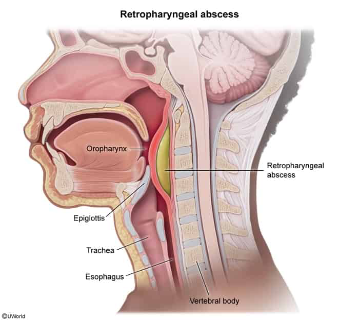
- High fever, severe sore throat, dysphagia, drooling, “hot potato” voice.
- Neck hyperextension (posturing for airway), pain w/ neck extension, bulging of posterior pharyngeal wall.
-
Diagnosis:
- Airway First: Secure if in respiratory distress.
- Initial Imaging: Lateral neck X-ray (widened retropharyngeal soft tissue: >7 mm at C2, >14 mm at C6 in kids).
- Confirmatory Imaging: CT neck w/ contrast (hypodense collection w/ rim enhancement).
-
Key Differential Diagnostics:
- Peritonsillar Abscess: Trismus, uvular deviation (contralateral), tonsillar swelling.
- Epiglottitis: Thumbprint sign, tripod position, cherry-red epiglottis.
- Croup: Barking cough, steeple sign (subglottic narrowing).
- Ludwig Angina: Submandibular space cellulitis, elevated tongue, firm mouth floor.
-
Management:
- Airway Protection: Emergent intubation/tracheostomy if stridor/distress.
- Empiric IV Abx: Ampicillin-sulbactam OR Ceftriaxone + Metronidazole (+ Vancomycin if MRSA suspected).
- Surgical I&D: Indicated if large (≥2 cm), airway compromise, or failed Abx therapy at 24–48 hrs.
-
High-Yield Complications:
- Acute Mediastinitis: Spread down “danger space” (posterior to retropharyngeal space) to posterior mediastinum.
- Airway obstruction, aspiration pneumonia (rupture), IJV thrombosis, sepsis.